Wenn Sie das Fenster schließen, wird Ihre Konfiguration nicht gespeichert, es sei denn, Sie haben Ihren Artikel in die Bestellung aufgenommen oder zu Ihren Favoriten hinzugefügt.
Klicken Sie auf OK, um das MILLIPLEX® MAP-Tool zu schließen oder auf Abbrechen, um zu Ihrer Auswahl zurückzukehren.
Wählen Sie konfigurierbare Panels & Premixed-Kits - ODER - Kits für die zelluläre Signaltransduktion & MAPmates™
Konfigurieren Sie Ihre MILLIPLEX® MAP-Kits und lassen sich den Preis anzeigen.
Konfigurierbare Panels & Premixed-Kits
Unser breites Angebot enthält Multiplex-Panels, für die Sie die Analyten auswählen können, die am besten für Ihre Anwendung geeignet sind. Unter einem separaten Register können Sie das Premixed-Cytokin-Format oder ein Singleplex-Kit wählen.
Kits für die zelluläre Signaltransduktion & MAPmates™
Wählen Sie gebrauchsfertige Kits zur Erforschung gesamter Signalwege oder Prozesse. Oder konfigurieren Sie Ihre eigenen Kits mit Singleplex MAPmates™.
Die folgenden MAPmates™ sollten nicht zusammen analysiert werden: -MAPmates™, die einen unterschiedlichen Assaypuffer erfordern. -Phosphospezifische und MAPmate™ Gesamtkombinationen wie Gesamt-GSK3β und Gesamt-GSK3β (Ser 9). -PanTyr und locusspezifische MAPmates™, z.B. Phospho-EGF-Rezeptor und Phospho-STAT1 (Tyr701). -Mehr als 1 Phospho-MAPmate™ für ein einziges Target (Akt, STAT3). -GAPDH und β-Tubulin können nicht mit Kits oder MAPmates™, die panTyr enthalten, analysiert werden.
.
Bestellnummer
Bestellinformationen
St./Pkg.
Liste
Dieser Artikel wurde zu Ihren Favoriten hinzugefügt.
Wählen Sie bitte Spezies, Panelart, Kit oder Probenart
Um Ihr MILLIPLEX® MAP-Kit zu konfigurieren, wählen Sie zunächst eine Spezies, eine Panelart und/oder ein Kit.
Custom Premix Selecting "Custom Premix" option means that all of the beads you have chosen will be premixed in manufacturing before the kit is sent to you.
Catalogue Number
Ordering Description
Qty/Pack
List
Dieser Artikel wurde zu Ihren Favoriten hinzugefügt.
Spezies
Panelart
Gewähltes Kit
Menge
Bestellnummer
Bestellinformationen
St./Pkg.
Listenpreis
96-Well Plate
Menge
Bestellnummer
Bestellinformationen
St./Pkg.
Listenpreis
Weitere Reagenzien hinzufügen (MAPmates erfordern die Verwendung eines Puffer- und Detektionskits)
Menge
Bestellnummer
Bestellinformationen
St./Pkg.
Listenpreis
48-602MAG
Buffer Detection Kit for Magnetic Beads
1 Kit
Platzsparende Option Kunden, die mehrere Kits kaufen, können ihre Multiplex-Assaykomponenten in Kunststoffbeuteln anstelle von Packungen erhalten, um eine kompaktere Lagerung zu ermöglichen.
Dieser Artikel wurde zu Ihren Favoriten hinzugefügt.
Das Produkt wurde in Ihre Bestellung aufgenommen
Sie können nun ein weiteres Kit konfigurieren, ein Premixed-Kit wählen, zur Kasse gehen oder das Bestell-Tool schließen.
Anti-CEMIP, clone PW-3, Cat. No. MABC1764, is a mouse monoclonal antibody that detects CEMIP and is tested for use in Immunohistochemistry and Western Blotting.
More>>Anti-CEMIP, clone PW-3, Cat. No. MABC1764, is a mouse monoclonal antibody that detects CEMIP and is tested for use in Immunohistochemistry and Western Blotting. Less<<
Empfohlene Produkte
Übersicht
Replacement Information
Description
Catalogue Number
MABC1764-25UG
Description
Anti-CEMIP Antibody, clone PW-3
Alternate Names
Cell migration-inducing and hyaluronan-binding protein
CCSP1
Background Information
Cell migration-inducing and hyaluronan-binding protein (UniProt: Q8WUJ3; also known as EC:3.2.1.35, CCSP1, CEMIP) is encoded by the CEMIP (also known as KIAA1199) gene (Gene ID: 57214) in human. CEMIP is highly glycosylated protein that is widely distributed in the cytoplasm and is localized in endoplasmic reticulum, endosomes, cell membrane, and in exosomes. It is also abundantly secreted. It is synthesized with a signal peptide (aa 1-30) that is essential for its proper translocation, hyaluronic acid (HA) degradation activity, and secretion. It mediates depolymerization of HA via the cell membrane-associated clathrin-coated pit endocytic pathway. It hydrolyzes high molecular weight HA to produce an intermediate-sized product, a process that may occur through rapid vesicle endocytosis and recycling without intracytoplasmic accumulation or digestion in lysosomes. CEMIP contains two GG domains, one G8 domain (aa 44-166), and four PbH1 domains (aa 572-819). The G8 domain contains eight conserved glycine residues and is reported to bind to Annexin 1 that enables it to adhere to cell membrane in Annexin 1 positive cells. The two GG domains are composed of seven β-strands and two helices and are located N-terminally after the G8 domain. Each of the four PbH1 domains contain 22 to 23 amino acids and function in polysaccharide hydrolysis. Amino acids sequence 295-591, also known as the B domain, is essential for its retention in the endoplasmic reticulum. CEMIP expression is strongly upregulated in many types of cancer and its higher levels are associated with poor prognosis in cancer patients. Higher expression of CEMIP is reported in ductal carcinoma and invasive breast cancer cells, gastric cancers, and in colon adenocarcinomas. It is considered as a potential blood-borne biomarker for colorectal cancers. The knockdown of CEMIP gene has been shown to significantly reduce metastasis to the lung, liver, and various other organs. (Ref.: Domanegg, K., et al (2022). Cancers (Basel). 14(20); 5093; Fink, SP., et al. (2015). Oncotarget, Vol. 6(31); 30500-30515).
References
Product Information
Format
Purified
Presentation
Purified mouse monoclonal antibody IgG1 in buffer containing 0.1 M Tris-Glycine (pH 7.4), 150 mM NaCl with 0.05% sodium azide.
Anti-CEMIP, clone PW-3, Cat. No. MABC1764, is a mouse monoclonal antibody that detects CEMIP and is tested for use in Immunohistochemistry and Western Blotting.
Key Applications
Western Blotting
Immunohistochemistry
Application Notes
Tested Applications
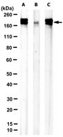
Western Blotting Analysis: A 1:1,000 dilution from a representative lot detected CEMIP in Caco-2 and HT29 cell lysates.
Immunohistochemistry Applications: A representative lot detected CEMIP, clone PW-3 in Immunohistochemistry applications (Fink, S.P., et al. (2015). Oncotarget. 6(31):30500-15).
Western Blotting Analysis: A representative lot detected CEMIP in Western Blotting applications (Fink, S.P., et al. (2015). Oncotarget. 6(31):30500-15).
Note: Actual optimal working dilutions must be determined by end user as specimens, and experimental conditions may vary with the end user.
Biological Information
Immunogen
V5/His-tagged, full-length, recombinant human Cell migration-inducing and hyaluronan-binding protein (CEMIP).
Epitope
Unknown
Clone
PW-3
Concentration
1 mg/mL. Please refer to guidance on suggested starting dilutions and/or titers per application and sample type.
Host
Mouse
Specificity
Clone PW-3 is a mouse monoclonal antibody that detects Cell migration-inducing and hyaluronan-binding protein (CEMIP).
180 kDa observed. 153.0 kDa calculated. Uncharacterized bands may be observed in some lysate(s).
Physicochemical Information
Dimensions
Materials Information
Toxicological Information
Safety Information according to GHS
Safety Information
Product Usage Statements
Quality Assurance
Evaluated by Western Blotting in DLD-1 cell lysate.
Western Blotting Analysis: A 1:1,000 dilution of this antibody detected CEMIP in DLD-1 cell lysate.
Usage Statement
Unless otherwise stated in our catalog or other company documentation accompanying the product(s), our products are intended for research use only and are not to be used for any other purpose, which includes but is not limited to, unauthorized commercial uses, in vitro diagnostic uses, ex vivo or in vivo therapeutic uses or any type of consumption or application to humans or animals.