Wenn Sie das Fenster schließen, wird Ihre Konfiguration nicht gespeichert, es sei denn, Sie haben Ihren Artikel in die Bestellung aufgenommen oder zu Ihren Favoriten hinzugefügt.
Klicken Sie auf OK, um das MILLIPLEX® MAP-Tool zu schließen oder auf Abbrechen, um zu Ihrer Auswahl zurückzukehren.
Wählen Sie konfigurierbare Panels & Premixed-Kits - ODER - Kits für die zelluläre Signaltransduktion & MAPmates™
Konfigurieren Sie Ihre MILLIPLEX® MAP-Kits und lassen sich den Preis anzeigen.
Konfigurierbare Panels & Premixed-Kits
Unser breites Angebot enthält Multiplex-Panels, für die Sie die Analyten auswählen können, die am besten für Ihre Anwendung geeignet sind. Unter einem separaten Register können Sie das Premixed-Cytokin-Format oder ein Singleplex-Kit wählen.
Kits für die zelluläre Signaltransduktion & MAPmates™
Wählen Sie gebrauchsfertige Kits zur Erforschung gesamter Signalwege oder Prozesse. Oder konfigurieren Sie Ihre eigenen Kits mit Singleplex MAPmates™.
Die folgenden MAPmates™ sollten nicht zusammen analysiert werden: -MAPmates™, die einen unterschiedlichen Assaypuffer erfordern. -Phosphospezifische und MAPmate™ Gesamtkombinationen wie Gesamt-GSK3β und Gesamt-GSK3β (Ser 9). -PanTyr und locusspezifische MAPmates™, z.B. Phospho-EGF-Rezeptor und Phospho-STAT1 (Tyr701). -Mehr als 1 Phospho-MAPmate™ für ein einziges Target (Akt, STAT3). -GAPDH und β-Tubulin können nicht mit Kits oder MAPmates™, die panTyr enthalten, analysiert werden.
.
Bestellnummer
Bestellinformationen
St./Pkg.
Liste
Dieser Artikel wurde zu Ihren Favoriten hinzugefügt.
Wählen Sie bitte Spezies, Panelart, Kit oder Probenart
Um Ihr MILLIPLEX® MAP-Kit zu konfigurieren, wählen Sie zunächst eine Spezies, eine Panelart und/oder ein Kit.
Custom Premix Selecting "Custom Premix" option means that all of the beads you have chosen will be premixed in manufacturing before the kit is sent to you.
Catalogue Number
Ordering Description
Qty/Pack
List
Dieser Artikel wurde zu Ihren Favoriten hinzugefügt.
Spezies
Panelart
Gewähltes Kit
Menge
Bestellnummer
Bestellinformationen
St./Pkg.
Listenpreis
96-Well Plate
Menge
Bestellnummer
Bestellinformationen
St./Pkg.
Listenpreis
Weitere Reagenzien hinzufügen (MAPmates erfordern die Verwendung eines Puffer- und Detektionskits)
Menge
Bestellnummer
Bestellinformationen
St./Pkg.
Listenpreis
48-602MAG
Buffer Detection Kit for Magnetic Beads
1 Kit
Platzsparende Option Kunden, die mehrere Kits kaufen, können ihre Multiplex-Assaykomponenten in Kunststoffbeuteln anstelle von Packungen erhalten, um eine kompaktere Lagerung zu ermöglichen.
Dieser Artikel wurde zu Ihren Favoriten hinzugefügt.
Das Produkt wurde in Ihre Bestellung aufgenommen
Sie können nun ein weiteres Kit konfigurieren, ein Premixed-Kit wählen, zur Kasse gehen oder das Bestell-Tool schließen.
AB5156-50UL
Sigma-AldrichAnti-Calcium Channel Antibody, Voltage Gated α 1C
Anti-Calcium Channel Antibody, Voltage Gated α 1C, detects levels of Voltage Gated α 1C Calcium Channel & has been published & validated for use in IH, IP & WB.
More>>Anti-Calcium Channel Antibody, Voltage Gated α 1C, detects levels of Voltage Gated α 1C Calcium Channel & has been published & validated for use in IH, IP & WB. Less<<
SDB (Sicherheitsdatenblätter), Analysenzertifikate und Qualitätszertifikate, Dossiers, Broschüren und andere verfügbare Dokumente.
Voltage-sensitive calcium channels (vscc) mediate the entry of calcium ions into excitable cells and are also involved in a variety of calcium-dependent processes, including muscle contraction, hormone or neurotransmitter release, gene expression, cell motility, cell division and cell death. The isoform alpha-1c gives rise to l-type calcium currents. Long-lasting (l-type) calcium channels belong to the "high-voltage activated" (hva) group. They are blocked by dihydropyridines (dhp), phenylalkylamines, benzothiazepines, and by omega-agatoxin-IIIA (omega-aga-IIIA). They are however insensitive to omega-conotoxin- gvia (omega-ctx-gvia) and omega-agatoxin-IVA (omega-aga-IVA). Calcium channels containing the alpha-1C subunit play an important role in excitation-contraction coupling in the heart. The various isoforms display marked differences in the sensitivity to dhp compounds (Swiss Prot).
References
Product Information
Format
Affinity Purified
Control
CONTROL ANTIGEN: Included free of charge with the antibody is 20 μg of control antigen (lyophilized powder in phosphate buffered saline, pH 7.4, containing 5% sucrose and 0.025% sodium azide). The stock solution of the antigen can be made up using 200 μL of sterile deionized water. For negative control, preincubate 1 μg of peptide with 1 μg of antibody for one hour at room temperature. Optimal concentrations must be determined by the end user.
Presentation
Affinity purified immunoglobulin. Lyophilized from phosphate buffered saline, pH 7.4, containing 1% BSA and 0.05% sodium azide as a preservative. Reconstitute with 50 μL of sterile deionized water. Centrifuge antibody preparation before use (10,000 xg for 5 min).
Anti-Calcium Channel Antibody, Voltage Gated α 1C, detects levels of Voltage Gated α 1C Calcium Channel & has been published & validated for use in IH, IP & WB.
Key Applications
Immunohistochemistry
Immunoprecipitation
Western Blotting
Application Notes
Alpha 1 subunits of voltage-gated Ca2+ channels are highly sensitive to proteases. All procedures that are going to receive a full-length protein should be performed at 4°C, and the following protease inhibitor mixture should be used: pepstatin A (1 μg/mL), leupeptin (1 μg/mL), aprotinin (1 μg/mL), Pefabloc SC (0.2 mM), benzamidine (0.1 mg/mL), and calpain inhibitors I and II (8 μg/mL each).
Immunohistochemistry: 1:200 on rat brain sections.
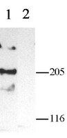
Western blot: 1:200 using ECL on rat brain and heart material. LMW form shows a 190kDa band while the HMW form shows a 210kDa band.
Immunoprecipitation
Dilutions should be made using a carrier protein such as BSA (1-3%)
Optimal working dilutions must be determined by the end user.
Biological Information
Immunogen
Purified peptide (C)TTKI NMDDL QPSEN EDKS (CNC1) from 848-865 of alpha 1C subunit of rat brain voltage-gated calcium channel (Accession number P22002). Epitope location is in the intracellular loop between domains II and III.
Concentration
Please refer to the Certificate of Analysis for the lot-specific concentration.
Host
Rabbit
Specificity
Recognizes a1C subunit (both LMW and HMW forms) of voltage-gated calcium channel. Does not cross react with any other calcium channel antigens tested so far. Guinea pig (17/18 residues identical); human and rabbit (16/18 residues identical).
This gene encodes an alpha-1 subunit of a voltage-dependent calcium channel. Calcium channels mediate the influx of calcium ions into the cell upon membrane polarization. The alpha-1 subunit consists of 24 transmembrane segments and forms the pore through which ions pass into the cell. The calcium channel consists of a complex of alpha-1, alpha-2/delta, beta, and gamma subunits in a 1:1:1:1 ratio. There are multiple isoforms of each of these proteins, either encoded by different genes or the result of alternative splicing of transcripts. The protein encoded by this gene binds to and is inhibited by dihydropyridine. Many alternate transcriptional splice variants of this gene have been observed but have not been thoroughly characterized.
FUNCTION: SwissProt: Q13936 # Voltage-sensitive calcium channels (VSCC) mediate the entry of calcium ions into excitable cells and are also involved in a variety of calcium-dependent processes, including muscle contraction, hormone or neurotransmitter release, gene expression, cell motility, cell division and cell death. The isoform alpha-1C gives rise to L-type calcium currents. Long-lasting (L-type) calcium channels belong to the high-voltage activated (HVA) group. They are blocked by dihydropyridines (DHP), phenylalkylamines, benzothiazepines, and by omega-agatoxin-IIIA (omega-Aga-IIIA). They are however insensitive to omega-conotoxin- GVIA (omega-CTx-GVIA) and omega-agatoxin-IVA (omega-Aga-IVA). Calcium channels containing the alpha-1C subunit play an important role in excitation-contraction coupling in the heart. The various isoforms display marked differences in the sensitivity to DHP compounds. SIZE: 2221 amino acids; 248887 Da SUBUNIT: Voltage-dependent calcium channels are multisubunit complexes, consisting of alpha-1, alpha-2, beta and delta subunits in a 1:1:1:1 ratio. The channel activity is directed by the pore- forming and voltage-sensitive alpha-1 subunit. In many cases, this subunit is sufficient to generate voltage-sensitive calcium channel activity. The auxiliary subunits beta and alpha-2/delta linked by a disulfide bridge regulate the channel activity. Interacts with CACNA2D4. SUBCELLULAR LOCATION: Membrane; Multi-pass membrane protein. TISSUE SPECIFICITY: Expressed in brain, heart, jejunum, ovary, pancreatic beta-cells and vascular smooth muscle. Overall expression is reduced in atherosclerotic vascular smooth muscle. DOMAIN: SwissProt: Q13936 Each of the four internal repeats contains five hydrophobic transmembrane segments (S1, S2, S3, S5, S6) and one positively charged transmembrane segment (S4). S4 segments probably represent the voltage-sensor and are characterized by a series of positively charged amino acids at every third position. & Binding of intracellular calcium through the EF-hand motif inhibits the opening of the channel (By similarity). PTM: Phosphorylation by PKA activates the channel (By similarity). DISEASE: SwissProt: Q13936 # Defects in CACNA1C are the cause of Timothy syndrome (TS) [MIM:601005]. TS is a disorder characterized by multiorgan dysfunction including lethal arrhythmias, webbing of fingers and toes, congenital heart disease, immune deficiency, intermittent hypoglycemia, cognitive abnormalities and autism. SIMILARITY: Belongs to the calcium channel alpha-1 subunit family.
Physicochemical Information
Dimensions
Materials Information
Toxicological Information
Safety Information according to GHS
Safety Information
Product Usage Statements
Usage Statement
Unless otherwise stated in our catalog or other company documentation accompanying the product(s), our products are intended for research use only and are not to be used for any other purpose, which includes but is not limited to, unauthorized commercial uses, in vitro diagnostic uses, ex vivo or in vivo therapeutic uses or any type of consumption or application to humans or animals.
Storage and Shipping Information
Storage Conditions
Maintain lyophilized material at -20°C for up to 12 months. After reconstitution add glycerol (ASC grade or better) at a ratio of 1:1 and maintain at -20°C for up to 6 months. Avoid repeated freeze/thaw cycles.
Packaging Information
Material Size
50 µL
Transport Information
Supplemental Information
Specifications
Global Trade ITEM Number
Bestellnummer
GTIN
AB5156-50UL
04053252373299
Documentation
Anti-Calcium Channel Antibody, Voltage Gated α 1C SDB
Signaling to the nucleus by an L-type calcium channel-calmodulin complex through the MAP kinase pathway. Dolmetsch, R E, et al. Science, 294: 333-9 (2001)
2001
Increases in the intracellular concentration of calcium ([Ca2+]i) activate various signaling pathways that lead to the expression of genes that are essential for dendritic development, neuronal survival, and synaptic plasticity. The mode of Ca2+ entry into a neuron plays a key role in determining which signaling pathways are activated and thus specifies the cellular response to Ca2+. Ca2+ influx through L-type voltage-activated channels (LTCs) is particularly effective at activating transcription factors such as CREB and MEF-2. We developed a functional knock-in technique to investigate the features of LTCs that specifically couple them to the signaling pathways that regulate gene expression. We found that an isoleucine-glutamine ("IQ") motif in the carboxyl terminus of the LTC that binds Ca2+-calmodulin (CaM) is critical for conveying the Ca2+ signal to the nucleus. Ca2+-CaM binding to the LTC was necessary for activation of the Ras/mitogen-activated protein kinase (MAPK) pathway, which conveys local Ca2+ signals from the mouth of the LTC to the nucleus. CaM functions as a local Ca2+ sensor at the mouth of the LTC that activates the MAPK pathway and leads to the stimulation of genes that are essential for neuronal survival and plasticity.