Wenn Sie das Fenster schließen, wird Ihre Konfiguration nicht gespeichert, es sei denn, Sie haben Ihren Artikel in die Bestellung aufgenommen oder zu Ihren Favoriten hinzugefügt.
Klicken Sie auf OK, um das MILLIPLEX® MAP-Tool zu schließen oder auf Abbrechen, um zu Ihrer Auswahl zurückzukehren.
Wählen Sie konfigurierbare Panels & Premixed-Kits - ODER - Kits für die zelluläre Signaltransduktion & MAPmates™
Konfigurieren Sie Ihre MILLIPLEX® MAP-Kits und lassen sich den Preis anzeigen.
Konfigurierbare Panels & Premixed-Kits
Unser breites Angebot enthält Multiplex-Panels, für die Sie die Analyten auswählen können, die am besten für Ihre Anwendung geeignet sind. Unter einem separaten Register können Sie das Premixed-Cytokin-Format oder ein Singleplex-Kit wählen.
Kits für die zelluläre Signaltransduktion & MAPmates™
Wählen Sie gebrauchsfertige Kits zur Erforschung gesamter Signalwege oder Prozesse. Oder konfigurieren Sie Ihre eigenen Kits mit Singleplex MAPmates™.
Die folgenden MAPmates™ sollten nicht zusammen analysiert werden: -MAPmates™, die einen unterschiedlichen Assaypuffer erfordern. -Phosphospezifische und MAPmate™ Gesamtkombinationen wie Gesamt-GSK3β und Gesamt-GSK3β (Ser 9). -PanTyr und locusspezifische MAPmates™, z.B. Phospho-EGF-Rezeptor und Phospho-STAT1 (Tyr701). -Mehr als 1 Phospho-MAPmate™ für ein einziges Target (Akt, STAT3). -GAPDH und β-Tubulin können nicht mit Kits oder MAPmates™, die panTyr enthalten, analysiert werden.
.
Bestellnummer
Bestellinformationen
St./Pkg.
Liste
Dieser Artikel wurde zu Ihren Favoriten hinzugefügt.
Wählen Sie bitte Spezies, Panelart, Kit oder Probenart
Um Ihr MILLIPLEX® MAP-Kit zu konfigurieren, wählen Sie zunächst eine Spezies, eine Panelart und/oder ein Kit.
Custom Premix Selecting "Custom Premix" option means that all of the beads you have chosen will be premixed in manufacturing before the kit is sent to you.
Catalogue Number
Ordering Description
Qty/Pack
List
Dieser Artikel wurde zu Ihren Favoriten hinzugefügt.
Spezies
Panelart
Gewähltes Kit
Menge
Bestellnummer
Bestellinformationen
St./Pkg.
Listenpreis
96-Well Plate
Menge
Bestellnummer
Bestellinformationen
St./Pkg.
Listenpreis
Weitere Reagenzien hinzufügen (MAPmates erfordern die Verwendung eines Puffer- und Detektionskits)
Menge
Bestellnummer
Bestellinformationen
St./Pkg.
Listenpreis
48-602MAG
Buffer Detection Kit for Magnetic Beads
1 Kit
Platzsparende Option Kunden, die mehrere Kits kaufen, können ihre Multiplex-Assaykomponenten in Kunststoffbeuteln anstelle von Packungen erhalten, um eine kompaktere Lagerung zu ermöglichen.
Dieser Artikel wurde zu Ihren Favoriten hinzugefügt.
Das Produkt wurde in Ihre Bestellung aufgenommen
Sie können nun ein weiteres Kit konfigurieren, ein Premixed-Kit wählen, zur Kasse gehen oder das Bestell-Tool schließen.
This Anti-Dual oxidase 2, clone Duox S-12 Antibody is validated for use in Western Blotting and Immunohistochemistry and Immunocytochemistry for the detection of Dual oxidase 2.
More>>This Anti-Dual oxidase 2, clone Duox S-12 Antibody is validated for use in Western Blotting and Immunohistochemistry and Immunocytochemistry for the detection of Dual oxidase 2. Less<<
SDB (Sicherheitsdatenblätter), Analysenzertifikate und Qualitätszertifikate, Dossiers, Broschüren und andere verfügbare Dokumente.
Dual oxidase 2 is an important enzyme in thyroid hormone biosynthesis and peroxide metabolism. Dual oxidase 2 generates the hydrogen peroxide needed for thyroid peroxidase and lactoperoxidase enzymes as well as processes H2O2 made during metabolic processes. Dual oxidase also plays a role in anti-microbial defense in our mucous membranes by providing the peroxidase for lactoperoxidase’s antimicrobial activities. Dual oxidase 2 is also needed for various extracellular matrix stabilizations and protein cross-linking needs such as during cuticle formation. Dual oxidase 2 is widely expressed in the developing fetus, and in the adult, it is largely expressed in the mucosal and epithelial cells and linings of the gut, lung, salivary glands and testis. Dual oxidase also plays a role in many hypersensitivity and inflammatory diseases and is upregulated in diseases such as colitis and certain cancers.
References
Product Information
Format
Purified
Presentation
Purified mouse monoclonal IgG1κ in buffer containing 0.1 M Tris-Glycine (pH 7.4), 150 mM NaCl with 0.05% sodium azide.
This Anti-Dual oxidase 2, clone Duox S-12 Antibody is validated for use in Western Blotting and Immunohistochemistry and Immunocytochemistry for the detection of Dual oxidase 2.
Key Applications
Western Blotting
Immunohistochemistry
Immunocytochemistry
Application Notes
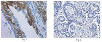
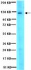
Immunohistochemistry Analysis: A 1:50 dilution from a representative lot detected Dual oxidase 2 in human prostate cancer and human breast cancer tissue. Western Blotting Analysis: A representative lot detected Dual oxidase 2 in BxPC-3 cell lysate (Wu, Y., et al. (2013). International Journal of Oncology. 42:1229-1238). Immunocytochemistry Analysis: A representative lot detected Dual oxidase 2 in MIA PACA-2 cell (Wu, Y., et al. (2013). International Journal of Oncology. 42:1229-1238). Immunohistochemistry Analysis: A representative lot detected Dual oxidase 2 MIA PACA-2 tissue (Wu, Y., et al. (2013). International Journal of Oncology. 42:1229-1238).
Biological Information
Immunogen
Recombinant protein corresponding to the N-terminus of human Dual oxidase 2.
Evaluated by Western Blotting in BxPC3 treated with IL-4 cell lysate.
Western Blotting Analysis: 1.0 µg/mL of this antibody detected Dual oxidase 2 in 10 µg of BxPC3 treated with IL-4 cell lysate.
Usage Statement
Unless otherwise stated in our catalog or other company documentation accompanying the product(s), our products are intended for research use only and are not to be used for any other purpose, which includes but is not limited to, unauthorized commercial uses, in vitro diagnostic uses, ex vivo or in vivo therapeutic uses or any type of consumption or application to humans or animals.
Functional activity and tumor-specific expression of dual oxidase 2 in pancreatic cancer cells and human malignancies characterized with a novel monoclonal antibody. Wu, Yonghzong, et al. Int. J. Oncol., 42: 1229-38 (2013)
2013
Dual oxidase 2 (Duox2), one of the seven members of the NADPH oxidase gene family, plays a critical role in generating H2O2 for thyroid hormone biosynthesis and as an integral part of the host defense system of the respiratory epithelium and the gastrointestinal tract. Recent evidence suggests that the regulation of Duox2 expression is under the control of pro-inflammatory cytokines and that Duox2-induced reactive oxygen species (ROS) contribute to the inflammation-related tissue injury that occurs in two pre-malignant, inflammatory conditions: chronic pancreatitis and inflammatory bowel disease. Because no reliable Duox antibodies are commercially available, we report the development of a murine monoclonal antibody (MAb) to Duox2 (clone Duox S-12) and its use for the characterization of Duox2 expression in human tumors, tumor cell lines and normal tissues. Duox S-12 specifically detected both endogenously- and ectopically-expressed Duox2 protein by immunoblotting, immunofluorescence microscopy and immunohistochemistry (where both membranous and cytoplasmic staining were present). Duox2 expression detected by Duox S-12 was functionally coupled to the generation of H(2)O(2) in pancreatic cancer cells that expressed Duox2 and its cognate maturation factor DuoxA2. Although Duox S-12 recognizes ectopically expressed Duox1 protein because of the extensive amino acid homology between Duox1 and Duox2, the lack of substantial Duox1 mRNA expression in human tumors (except thyroid cancer) allowed us to evaluate Duox2 expression across a wide range of normal and malignant tissues by immuno-histochemistry. Duox2 was expressed at elevated levels in many human cancers, most notably tumors of the prostate, lung, colon and breast while brain tumors and lymphomas demonstrated the lowest frequency of expression. The Duox-specific monoclonal antibody described here provides a promising tool for the further examination of the role of Duox-dependent reactive oxygen production in inflammation-related carcinogenesis, where alterations in oxidant tone play a critical role in cell growth and proliferation.