Wenn Sie das Fenster schließen, wird Ihre Konfiguration nicht gespeichert, es sei denn, Sie haben Ihren Artikel in die Bestellung aufgenommen oder zu Ihren Favoriten hinzugefügt.
Klicken Sie auf OK, um das MILLIPLEX® MAP-Tool zu schließen oder auf Abbrechen, um zu Ihrer Auswahl zurückzukehren.
Wählen Sie konfigurierbare Panels & Premixed-Kits - ODER - Kits für die zelluläre Signaltransduktion & MAPmates™
Konfigurieren Sie Ihre MILLIPLEX® MAP-Kits und lassen sich den Preis anzeigen.
Konfigurierbare Panels & Premixed-Kits
Unser breites Angebot enthält Multiplex-Panels, für die Sie die Analyten auswählen können, die am besten für Ihre Anwendung geeignet sind. Unter einem separaten Register können Sie das Premixed-Cytokin-Format oder ein Singleplex-Kit wählen.
Kits für die zelluläre Signaltransduktion & MAPmates™
Wählen Sie gebrauchsfertige Kits zur Erforschung gesamter Signalwege oder Prozesse. Oder konfigurieren Sie Ihre eigenen Kits mit Singleplex MAPmates™.
Die folgenden MAPmates™ sollten nicht zusammen analysiert werden: -MAPmates™, die einen unterschiedlichen Assaypuffer erfordern. -Phosphospezifische und MAPmate™ Gesamtkombinationen wie Gesamt-GSK3β und Gesamt-GSK3β (Ser 9). -PanTyr und locusspezifische MAPmates™, z.B. Phospho-EGF-Rezeptor und Phospho-STAT1 (Tyr701). -Mehr als 1 Phospho-MAPmate™ für ein einziges Target (Akt, STAT3). -GAPDH und β-Tubulin können nicht mit Kits oder MAPmates™, die panTyr enthalten, analysiert werden.
.
Bestellnummer
Bestellinformationen
St./Pkg.
Liste
Dieser Artikel wurde zu Ihren Favoriten hinzugefügt.
Wählen Sie bitte Spezies, Panelart, Kit oder Probenart
Um Ihr MILLIPLEX® MAP-Kit zu konfigurieren, wählen Sie zunächst eine Spezies, eine Panelart und/oder ein Kit.
Custom Premix Selecting "Custom Premix" option means that all of the beads you have chosen will be premixed in manufacturing before the kit is sent to you.
Catalogue Number
Ordering Description
Qty/Pack
List
Dieser Artikel wurde zu Ihren Favoriten hinzugefügt.
Spezies
Panelart
Gewähltes Kit
Menge
Bestellnummer
Bestellinformationen
St./Pkg.
Listenpreis
96-Well Plate
Menge
Bestellnummer
Bestellinformationen
St./Pkg.
Listenpreis
Weitere Reagenzien hinzufügen (MAPmates erfordern die Verwendung eines Puffer- und Detektionskits)
Menge
Bestellnummer
Bestellinformationen
St./Pkg.
Listenpreis
48-602MAG
Buffer Detection Kit for Magnetic Beads
1 Kit
Platzsparende Option Kunden, die mehrere Kits kaufen, können ihre Multiplex-Assaykomponenten in Kunststoffbeuteln anstelle von Packungen erhalten, um eine kompaktere Lagerung zu ermöglichen.
Dieser Artikel wurde zu Ihren Favoriten hinzugefügt.
Das Produkt wurde in Ihre Bestellung aufgenommen
Sie können nun ein weiteres Kit konfigurieren, ein Premixed-Kit wählen, zur Kasse gehen oder das Bestell-Tool schließen.
07-130
Sigma-AldrichAnti-Ezrin Antibody
This Anti-Ezrin Antibody is validated for use in WB, IH for the detection of Ezrin.
More>>This Anti-Ezrin Antibody is validated for use in WB, IH for the detection of Ezrin. Less<<
Anti-Ezrin Antibody: SDB (Sicherheitsdatenblätter), Analysenzertifikate und Qualitätszertifikate, Dossiers, Broschüren und andere verfügbare Dokumente.
The cytoplasmic peripheral membrane protein encoded by this gene functions as a protein-tyrosine kinase substrate in microvilli. As a member of the ERM protein family, this protein serves as an intermediate between the plasma membrane and the actin cytoskeleton. This protein plays a key role in cell surface structure adhesion, migration and organization, and it has been implicated in various human cancers. A pseudogene located on chromosome 3 has been identified for this gene. Alternatively spliced variants have also been described for this gene.
FUNCTION: SwissProt: P15311 # Probably involved in connections of major cytoskeletal structures to the plasma membrane. SIZE: 586 amino acids; 69413 Da SUBUNIT: Interacts with MPP5 (By similarity). Interacts with SLC9A3R1 and PACE1. SUBCELLULAR LOCATION: Membrane. Note=Localization to the apical membrane of parietal cells depends on the interaction with MPP5. Localizes to cell extensions and peripheral processes of astrocytes (By similarity). Microvillar peripheral membrane protein (cytoplasmic side). TISSUE SPECIFICITY: Expressed in cerebral cortex, basal ganglia, hippocampus, hypophysis, and optic nerve. Weakly expressed in brain stem and diencephalon. Stronger expression was detected in gray matter of frontal lobe compared to white matter (at protein level). Component of the microvilli of intestinal epithelial cells. Preferentially expressed in astrocytes of hippocampus, frontal cortex, thalamus, parahippocampal cortex, amygdala, insula, and corpus callosum. Not detected in neurons in most tissues studied.DEVELOPMENTAL STAGE: Very strong staining is detected in the Purkinje cell layer and in part of the molecular layer of the infant brain compared to adult brain. PTM: Phosphorylated by tyrosine-protein kinases. SIMILARITY: SwissProt: P15311 ## Contains 1 FERM domain.
Molecular Weight
81kDa
Physicochemical Information
Dimensions
Materials Information
Toxicological Information
Safety Information according to GHS
Safety Information
Product Usage Statements
Quality Assurance
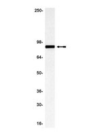
routinely evaluated by immunoblot on RIPA lysates from A431 cells or a mouse brain lysate
Usage Statement
Unless otherwise stated in our catalog or other company documentation accompanying the product(s), our products are intended for research use only and are not to be used for any other purpose, which includes but is not limited to, unauthorized commercial uses, in vitro diagnostic uses, ex vivo or in vivo therapeutic uses or any type of consumption or application to humans or animals.
Several organs, including the lungs and kidneys, are formed by epithelial tubes whose proper morphogenesis ensures correct function. This is best exemplified by the kidney, where defective establishment or maintenance of tubular diameter results in polycystic kidney disease, a common genetic disorder. Most polycystic kidney disease cases result from loss-of-function mutations in the PKD1 gene, encoding Polycystin-1, a large receptor of unknown function. Here we demonstrate that PC-1 has an essential role in the establishment of correct tubular diameter during nephron development. Polycystin-1 associates with Par3 favouring the assembly of a pro-polarizing Par3/aPKC complex and it regulates a programme of cell polarity important for oriented cell migration and for a convergent extension-like process during tubular morphogenesis. Par3 inactivation in the developing kidney results in defective convergent extension and tubular morphogenesis, and in renal cyst formation. Our data define Polycystin-1 as central to cell polarization and to epithelial tube morphogenesis and homeostasis.
Proteomics analysis of the ezrin interactome in B cells reveals a novel association with Myo18a?. Ken Matsui,Neetha Parameswaran,Nayer Bagheri,Belinda Willard,Neetu Gupta Journal of proteome research
10
2010
The molecular regulation of recruitment and assembly of signalosomes near the B cell receptor (BCR) is poorly understood. We have previously demonstrated a role for the ERM family protein ezrin in regulating antigen-dependent lipid raft coalescence in B cells. In this study, we addressed the possibility that ezrin may collaborate with other adaptor proteins to regulate signalosome dynamics at the membrane. Using mass spectrometry-based proteomics analysis, we identified Myo18a? as a novel binding partner of ezrin. Myo18a? is an attractive candidate as it has several protein-protein interaction domains and an intrinsic motor activity. The expression of Myo18a? varied during B cell development in the bone marrow and in mature B cell subsets suggesting functional differences. Interestingly, BCR stimulation increased the association between ezrin and Myo18a?, and induced co-segregation of Myo18a? with the BCR and phosphotyrosine-containing proteins. Our data raise an intriguing possibility that the Myo18a?/ezrin complex may facilitate BCR-mediated signaling by recruiting signaling proteins that are in close proximity of the antigen receptor. Our study is not only significant with respect to understanding the molecular regulation of BCR signaling but also provides a broader basis for understanding the mechanism of action of ezrin in other cellular systems.
Thyroid hormone receptors (TRs) mediate the critical activities of the thyroid hormone (T3) in growth, development, and differentiation. Decreased expression and/or somatic mutations of TRs have been shown to be associated with several types of human cancers including liver, breast, lung, and thyroid. A direct demonstration that TRβ mutants could function as oncogenes is evidenced by the spontaneous development of follicular thyroid carcinoma similar to human cancer in a knockin mouse model harboring a mutated TRβ (denoted as PV; Thrb(PV/PV) mice). PV is a dominant negative mutation identified in a patient with resistance to thyroid hormone. Analysis of altered gene expression and molecular studies of thyroid carcinogenesis in Thrb(PV/PV) mice show that the oncogenic activity of PV is mediated by both nucleus-initiated transcription and extranuclear actions to alter gene expression and signaling transduction activity. This article focuses on recent findings of novel extranuclear actions of PV that affect signaling cascades and thereby the invasiveness, migration, and motility of thyroid tumor cells. These findings have led to identification of potential molecular targets for treatment of metastatic thyroid cancer.
Differentiation is the process by which tissues/organs take on their final, physiologically functional form. This process is mediated in part by the silencing of embryonic genes and the activation of terminal, differentiation gene products. Mammalian kidney development is initiated when the Wolffian duct branches and invades the overlying metanephric mesenchyme. The newly formed epithelial bud, known as the ureteric bud, will continue to branch ultimately differentiating into the collecting duct system and ureter. Here, we show that Hoxb7-Cre mediated removal of beta-catenin from the mouse Wolffian duct epithelium leads to the premature expression of gene products normally associated with the differentiated kidney collecting duct system including the water channel protein, Aquaporin-3 and the tight junction protein isoform, ZO-1 alpha+. Mutant cells fail to maintain expression of some genes associated with embryonic development, including several mediators of branching morphogenesis, which subsequently leads to kidney aplasia or hypoplasia. Reciprocally, expression of a stabilized form of beta-catenin appears to block differentiation of the collecting ducts. All of these defects occur in the absence of any effects on the adherens junctions. These data indicate a role for beta-catenin in maintaining cells of the Wolffian ducts and the duct derived ureteric bud/collecting duct system in an undifferentiated or precursor state.
Myc down-regulation as a mechanism to activate the Rb pathway in STAT5A-induced senescence. Mallette, FA; Gaumont-Leclerc, MF; Huot, G; Ferbeyre, G The Journal of biological chemistry
282
34938-44
2007
Senescence is a general antiproliferative program that avoids the expansion of cells bearing oncogenic mutations. We found that constitutively active STAT5A (ca-STAT5A) can induce a p53- and Rb-dependent cellular senescence response. However, ca-STAT5A did not induce p21 and p16(INK4a), which are responsible for inhibiting cyclin-dependent protein kinases and engaging the Rb pathway during the senescence response to oncogenic ras. Intriguingly, ca-STAT5A led to a down-regulation of Myc and Myc targets, including CDK4, a negative regulator of Rb. The down-regulation of Myc was in part proteasome-dependent and correlated with its localization to promyelocytic leukemia bodies, which were found to be highly abundant during STAT5-induced senescence. Introduction of CDK4 or Myc bypassed STAT5A-induced senescence in cells in which p53 was also inactivated. These results uncover a novel mechanism to engage the Rb pathway in oncogene-induced senescence and indicate the existence of oncogene-specific pathways that regulate senescence.
Calpain regulates enterocyte brush border actin assembly and pathogenic Escherichia coli-mediated effacement Potter, D. A., et al J Biol Chem, 278:30403-12 (2003)
2003
Increased ezrin expression and activation by CDK5 coincident with acquisition of the senescent phenotype Yang, H. S. and Hinds, P. W. Mol Cell, 11:1163-76 (2003)
2003