Wenn Sie das Fenster schließen, wird Ihre Konfiguration nicht gespeichert, es sei denn, Sie haben Ihren Artikel in die Bestellung aufgenommen oder zu Ihren Favoriten hinzugefügt.
Klicken Sie auf OK, um das MILLIPLEX® MAP-Tool zu schließen oder auf Abbrechen, um zu Ihrer Auswahl zurückzukehren.
Wählen Sie konfigurierbare Panels & Premixed-Kits - ODER - Kits für die zelluläre Signaltransduktion & MAPmates™
Konfigurieren Sie Ihre MILLIPLEX® MAP-Kits und lassen sich den Preis anzeigen.
Konfigurierbare Panels & Premixed-Kits
Unser breites Angebot enthält Multiplex-Panels, für die Sie die Analyten auswählen können, die am besten für Ihre Anwendung geeignet sind. Unter einem separaten Register können Sie das Premixed-Cytokin-Format oder ein Singleplex-Kit wählen.
Kits für die zelluläre Signaltransduktion & MAPmates™
Wählen Sie gebrauchsfertige Kits zur Erforschung gesamter Signalwege oder Prozesse. Oder konfigurieren Sie Ihre eigenen Kits mit Singleplex MAPmates™.
Die folgenden MAPmates™ sollten nicht zusammen analysiert werden: -MAPmates™, die einen unterschiedlichen Assaypuffer erfordern. -Phosphospezifische und MAPmate™ Gesamtkombinationen wie Gesamt-GSK3β und Gesamt-GSK3β (Ser 9). -PanTyr und locusspezifische MAPmates™, z.B. Phospho-EGF-Rezeptor und Phospho-STAT1 (Tyr701). -Mehr als 1 Phospho-MAPmate™ für ein einziges Target (Akt, STAT3). -GAPDH und β-Tubulin können nicht mit Kits oder MAPmates™, die panTyr enthalten, analysiert werden.
.
Bestellnummer
Bestellinformationen
St./Pkg.
Liste
Dieser Artikel wurde zu Ihren Favoriten hinzugefügt.
Wählen Sie bitte Spezies, Panelart, Kit oder Probenart
Um Ihr MILLIPLEX® MAP-Kit zu konfigurieren, wählen Sie zunächst eine Spezies, eine Panelart und/oder ein Kit.
Custom Premix Selecting "Custom Premix" option means that all of the beads you have chosen will be premixed in manufacturing before the kit is sent to you.
Catalogue Number
Ordering Description
Qty/Pack
List
Dieser Artikel wurde zu Ihren Favoriten hinzugefügt.
Spezies
Panelart
Gewähltes Kit
Menge
Bestellnummer
Bestellinformationen
St./Pkg.
Listenpreis
96-Well Plate
Menge
Bestellnummer
Bestellinformationen
St./Pkg.
Listenpreis
Weitere Reagenzien hinzufügen (MAPmates erfordern die Verwendung eines Puffer- und Detektionskits)
Menge
Bestellnummer
Bestellinformationen
St./Pkg.
Listenpreis
48-602MAG
Buffer Detection Kit for Magnetic Beads
1 Kit
Platzsparende Option Kunden, die mehrere Kits kaufen, können ihre Multiplex-Assaykomponenten in Kunststoffbeuteln anstelle von Packungen erhalten, um eine kompaktere Lagerung zu ermöglichen.
Dieser Artikel wurde zu Ihren Favoriten hinzugefügt.
Das Produkt wurde in Ihre Bestellung aufgenommen
Sie können nun ein weiteres Kit konfigurieren, ein Premixed-Kit wählen, zur Kasse gehen oder das Bestell-Tool schließen.
This Anti-FoxA2/HNF-3b, clone 4C7, Cat. No. MABN2728, is a mouse monoclonal antibody that detects FoxA2/HGN-3b and is tested for use in EMSA, Immunocytochemistry, Immunofluorescence, Immunohistochemistry, Immunoprecipitation, and Western Blotting.
More>>This Anti-FoxA2/HNF-3b, clone 4C7, Cat. No. MABN2728, is a mouse monoclonal antibody that detects FoxA2/HGN-3b and is tested for use in EMSA, Immunocytochemistry, Immunofluorescence, Immunohistochemistry, Immunoprecipitation, and Western Blotting. Less<<
Empfohlene Produkte
Übersicht
Replacement Information
Description
Catalogue Number
MABN2728-100UL
Description
Anti-FoxA2/HNF-3b Antibody, clone 4C7
Alternate Names
Hepatocyte nuclear factor-3beta
HNF-3 beta
FOXA2
Forkhead box A2
Background Information
Hepatocyte nuclear factor-3beta (UniProt: Q9PWP8; also known as HNF-3 beta, FOXA2) is encoded by the HNF-3beta gene (Gene ID: 395539) in Chicken. FoxA2 belongs to the forkhead family of transcription factors that uses a modified helix-turn-helix motif to bind DNA as a monomer. Its DNA binding domain is localized in amino acids 151-245. It is expressed very early during development and is involved in the development of several tissues, including liver, pancreas, lung, prostate, and the nervous system. In mouse embryo, FoxA2 expression is initiated during gastrulation in the node, notochord, and floor plate. Its induction in neural plate cells is an early response to notochord-derived signals and its ectopic expression induces floor plate differentiation. In the ventral mesencephalon and cerebellum, it is co-expressed with Engrailed homeoproteins - En1 and En2, which raises the possibility that the two transcription factors interact to regulate common target genes. FoxA2 expression is observed in different regions of the developing nervous system. Along with FoxA1, it is reported to be crucial for the specification and differentiation of dopamine neurons during embryonic development. FoxA2 also functions as a suppressor of tumor metastasis by inhibition of epithelial-to-mesenchymal transition in human lung cancers. It is shown to be a key target of TGF-β1 in controlling epithelial-to-mesenchymal transition (EMT) in lung cancer cells. (Ref.: Rausa, FM., et al. (2020). Mol. Cell. Biol. 20(21); 8264-8282; Domanskyi, A., et al (2014). Front. Cell. Neurosci. 8; 275; Tang, Y., et al. (2011). Cell Res. 21(2); 316-326; Foucher, I., et al. (2003). Development 130(9); 1867-1876; Ericson, J., et al. (1996). Cell. 87(4); 661-673).
References
Product Information
Format
Purified
Presentation
Purified mouse monoclonal antibody IgG1 in buffer containing 0.1 M Tris-Glycine (pH 7.4), 150 mM NaCl with 0.05% sodium azide.
This Anti-FoxA2/HNF-3b, clone 4C7, Cat. No. MABN2728, is a mouse monoclonal antibody that detects FoxA2/HGN-3b and is tested for use in EMSA, Immunocytochemistry, Immunofluorescence, Immunohistochemistry, Immunoprecipitation, and Western Blotting.
Key Applications
Electrophoretic Mobility Shift Assay
Immunocytochemistry
Immunofluorescence
Immunohistochemistry
Immunoprecipitation
Western Blotting
Application Notes
Tested Applications
Electrophoretic Mobility Shift Assay: A representative lot detected FoxA2/HNF-3b in Electrophoretic Mobility Shift Assay (EMSA) (Foucher, I., et al. (2003). Development. 130(9): 1867-76).
Immunofluorescence Analysis: A representative lot detected FoxA2/HNF-3b in Immunofluorescence applications (Camp, D., et al. (2014). Development. 141(20): 3879-88; Hoelzl, M.A., et al. (2017). Dev Biol. 429(1): 132-146).
Immunoprecipitation Analysis: A representative lot immunoprecipitated FoxA2/HNF-3b in Immunoprecipitation applications (van den Brink, G.R., et al. (2001). Gastroenterology. 121(2):317-28).
Immunohistochemistry Applications: A representative lot detected FoxA2/HNF-3b in Immunohistochemistry applications (Ericson, J., et al. (1996). Cell. 87(4):661-73; Kwon, O., et al. (1998). J Clin Invest. 101(10):2054-64; Rousa, F.M., et al. (2000). Mol Cell Biol. 20(21):8264-82; van den Brink, G.R., et al. (2001). Gastroenterology. 121(2):317-28; Camp, D., et al. (2014). Development. 141(20):3879-88; Hoelzl, M.A., et al. (2017). Dev Biol. 429(1):132-146; Amarnath, S., et al. (2017). J Cell Sci. 130(1):119-131).
Immunocytochemistry Analysis: A representative lot detected FoxA2/HNF-3b in Immunohistochemistry applications (Nagase, T., et al. (2009). Dev Dyn. 238(5):1118-30).
Western Blotting Analysis: A representative lot detected FoxA2/HNF-3b in Western Blotting applications (Rousa, F.M., et al. (2000). Mol Cell Biol. 20(21):8264-82; van den Brink, G.R., et al. (2001). Gastroenterology. 121(2):317-28).
Note: Actual optimal working dilutions must be determined by end user as specimens, and experimental conditions may vary with the end user.
Biological Information
Immunogen
Recombinant Chicken Rhodopsin (1-6).
Epitope
Unknown
Clone
4C7
Concentration
0.5 mg/mL. Please refer to guidance on suggested starting dilutions and/or titers per application and sample type.
Host
Mouse
Specificity
Clone 4C7 is a mouse monoclonal antibody that detects Hepatocyte nuclear factor-3β (HNF-3β/FoxA2).
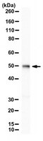
~50 kDa observed; 47.23 kDa calculated. Uncharacterized bands may be observed in some lysate(s).
Physicochemical Information
Dimensions
Materials Information
Toxicological Information
Safety Information according to GHS
Safety Information
Product Usage Statements
Quality Assurance
Evaluated by Western Blotting in HepG2 cell lysate.
Western Blotting Analysis: A 1:500 dilution of this antibody detected FoxA2/HNF-3b, in HepG2 cell lysate.
Usage Statement
Unless otherwise stated in our catalog or other company documentation accompanying the product(s), our products are intended for research use only and are not to be used for any other purpose, which includes but is not limited to, unauthorized commercial uses, in vitro diagnostic uses, ex vivo or in vivo therapeutic uses or any type of consumption or application to humans or animals.