Wenn Sie das Fenster schließen, wird Ihre Konfiguration nicht gespeichert, es sei denn, Sie haben Ihren Artikel in die Bestellung aufgenommen oder zu Ihren Favoriten hinzugefügt.
Klicken Sie auf OK, um das MILLIPLEX® MAP-Tool zu schließen oder auf Abbrechen, um zu Ihrer Auswahl zurückzukehren.
Wählen Sie konfigurierbare Panels & Premixed-Kits - ODER - Kits für die zelluläre Signaltransduktion & MAPmates™
Konfigurieren Sie Ihre MILLIPLEX® MAP-Kits und lassen sich den Preis anzeigen.
Konfigurierbare Panels & Premixed-Kits
Unser breites Angebot enthält Multiplex-Panels, für die Sie die Analyten auswählen können, die am besten für Ihre Anwendung geeignet sind. Unter einem separaten Register können Sie das Premixed-Cytokin-Format oder ein Singleplex-Kit wählen.
Kits für die zelluläre Signaltransduktion & MAPmates™
Wählen Sie gebrauchsfertige Kits zur Erforschung gesamter Signalwege oder Prozesse. Oder konfigurieren Sie Ihre eigenen Kits mit Singleplex MAPmates™.
Die folgenden MAPmates™ sollten nicht zusammen analysiert werden: -MAPmates™, die einen unterschiedlichen Assaypuffer erfordern. -Phosphospezifische und MAPmate™ Gesamtkombinationen wie Gesamt-GSK3β und Gesamt-GSK3β (Ser 9). -PanTyr und locusspezifische MAPmates™, z.B. Phospho-EGF-Rezeptor und Phospho-STAT1 (Tyr701). -Mehr als 1 Phospho-MAPmate™ für ein einziges Target (Akt, STAT3). -GAPDH und β-Tubulin können nicht mit Kits oder MAPmates™, die panTyr enthalten, analysiert werden.
.
Bestellnummer
Bestellinformationen
St./Pkg.
Liste
Dieser Artikel wurde zu Ihren Favoriten hinzugefügt.
Wählen Sie bitte Spezies, Panelart, Kit oder Probenart
Um Ihr MILLIPLEX® MAP-Kit zu konfigurieren, wählen Sie zunächst eine Spezies, eine Panelart und/oder ein Kit.
Custom Premix Selecting "Custom Premix" option means that all of the beads you have chosen will be premixed in manufacturing before the kit is sent to you.
Catalogue Number
Ordering Description
Qty/Pack
List
Dieser Artikel wurde zu Ihren Favoriten hinzugefügt.
Spezies
Panelart
Gewähltes Kit
Menge
Bestellnummer
Bestellinformationen
St./Pkg.
Listenpreis
96-Well Plate
Menge
Bestellnummer
Bestellinformationen
St./Pkg.
Listenpreis
Weitere Reagenzien hinzufügen (MAPmates erfordern die Verwendung eines Puffer- und Detektionskits)
Menge
Bestellnummer
Bestellinformationen
St./Pkg.
Listenpreis
48-602MAG
Buffer Detection Kit for Magnetic Beads
1 Kit
Platzsparende Option Kunden, die mehrere Kits kaufen, können ihre Multiplex-Assaykomponenten in Kunststoffbeuteln anstelle von Packungen erhalten, um eine kompaktere Lagerung zu ermöglichen.
Dieser Artikel wurde zu Ihren Favoriten hinzugefügt.
Das Produkt wurde in Ihre Bestellung aufgenommen
Sie können nun ein weiteres Kit konfigurieren, ein Premixed-Kit wählen, zur Kasse gehen oder das Bestell-Tool schließen.
MABN1783
Sigma-AldrichAnti-HLA Class I Antigens Antibody, clone W6/32
Anti-HLA Class I Antigens Antibody, clone W6/32, Cat. No. MABN1783, targets HLA class I antigens and has been tested in Affinity Binding, Flow Cytometry, Immunoaffinity Purification, Immunohistochemistry, Immunoprecipitation, and Neutralization applications.
More>>Anti-HLA Class I Antigens Antibody, clone W6/32, Cat. No. MABN1783, targets HLA class I antigens and has been tested in Affinity Binding, Flow Cytometry, Immunoaffinity Purification, Immunohistochemistry, Immunoprecipitation, and Neutralization applications. Less<<
SDB (Sicherheitsdatenblätter), Analysenzertifikate und Qualitätszertifikate, Dossiers, Broschüren und andere verfügbare Dokumente.
HLA class I histocompatibility antigen, A alpha chain
MHC class I antigen A
HLA class I histocompatibility antigen, B alpha chain
MHC class I antigen B
HLA class I histocompatibility antigen, C alpha chain
MHC class I antigen C;HLA class I histocompatibility antigen, E alpha chain
MHC class I antigen E
Background Information
Major histocompatibility complexes (MHCs), also called human leukocyte antigens (HLAs) in human, are cell surface molecules that play a major part of the immune system in all vertebrates by determining histocompatibility. The main function of MHCs is to bind and present peptide fragments derived from pathogens on the surface of antigen presenting cells. MHCs are divided into three classes, class I MHCs are heterodimers composed of a transmembrane alpha chain and a common extracellular beta subunit. The alpha chain contains three domains (alpha1, alpha2, and alpha3), with the alpha1 domain mediating interaction with the beta2 subunit. The alpha1 and alpha2 domains form the antigen-binding groove. Class II MHCs are heterodimers of two single transmembrane chains, alpha and beta, each contains two domains, with the N-terminal alpha1 and beta1 domain from each chain forming the antigen-binding groove. Class III molecules include several secreted proteins with immune functions, including components of the complement system, cytokines, and heat shock proteins. Each human cell expresses six MHC class I alleles (one HLA-A, -B, and -C allele from each parent). Classical MHCs present antigens to the TCRs of CD8+ T lymphocytes. Nonclassical molecules (class IB MHC) exhibit limited polymorphism, expression patterns, and presented antigens. This group is subdivided into a group encoded within MHC loci (e.g. HLA-E, -F, -G) and those not (e.g. ULBPs, Rae1, and H60).
References
Product Information
Format
Purified
Presentation
Purified mouse monoclonal antibody IgG2a in PBS without preservatives.
Anti-HLA Class I Antigens Antibody, clone W6/32, Cat. No. MABN1783, targets HLA class I antigens and has been tested in Affinity Binding, Flow Cytometry, Immunoaffinity Purification, Immunohistochemistry, Immunoprecipitation, and Neutralization applications.
Key Applications
Immunohistochemistry
Flow Cytometry
Immunoaffinity Purification
Neutralizing
Immunoprecipitation
Affinity Binding Assay
Application Notes
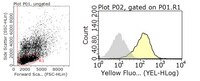
Flow Cytometry Analysis: 0.2 µL from a representative lot detected surface HLA class I antigens among the gated lymphocyte population from one million human PMBCs.
Affinity Binding Assay: A representative lot detected cell surface murine, rat, rabbit and guinea pig class I heavy chains upon association with bovine β2-microglobulin (β2-m), but not when associated with their respective autologous β2-m (Kahn-Perles, B., et al. (1987). J. Immunol. 138(7).2190-2196).
Affinity Binding Assay: A representative lot determined the level of HLA class I (A, B, C) antigens on the surface of human B cell lines and peripheral blood lymphocytes (Parham, P., et al. (1979). J. Immunol. 123(1): 342-349).
Affinity Binding Assay: A representative lot, pre-coupled to Sepharose beads, bound isolated HLA-A2 chain, but not isolated β2-microglobulin (β2-m). Pre-incubating HLA-A2 chain with β2-m enhanced the binding of HLA-A2 by clone W6/32 (Parham, P., et al. (1979). J. Immunol. 123(1): 342-349).
Flow Cytometry Analysis: A representative lot detected an inducction of HLA class I antigen expression on the surface of ESTDAB-004 and ESTDAB-159 human melanoma cells following IFN-α, but not IFN-γ, treatment (Rodríguez, T., et al. (2007). BMC Cancer. 7:34).
Flow Cytometry Analysis: Representative lots detected endogenous HLA-E on the surface of LCL721.221 human B lymphoblastoid cells, as well as exogenously expressed HLA-B on the surface of transfected LCL721.221 cells (Wooden, S.L., et al. (2005). J. Immunol. 175(3):1383-1387; Braud, V.M., et al. (1998). Curr. Biol. 8(1):1-10).
Flow Cytometry Analysis: A representative lot detected the restoration of HLA class I antigen expression on the surface of UKRV-Mel-2b human melanoma cells following β2-microglobulin (β2-m) transfection (Paschen, A., et al. (2003). Int. J. Cancer. 103(6):759-767).
Flow Cytometry Analysis: A representative lot detected HLA class I antigens on the surface of HEK293 cells. Surface HLA class I antigens level decreased upon exogenous expression of human adenovirus (Ad) E3/19K protein (Deryckere, F., and Burgert, H.G. (1996). J. Virol. 7(5):2832 2841).
Immunoaffinity Purification: A representative lot was coupled to Sepharose beads and affinity purified soluble HLA polypeptides of ~34/12 kDa from papain-digested crude membrane preparations of Bri 8 cells that express HLA-A, -B, -C, -D antigens, but not from Daudi cells that only express HLA-D antigens (Parham, P., et al. (1979). J. Immunol. 123(1): 342-349).
Immunohistochemistry Analysis: Representative lots detected HLA class I antigen-positive malignant tumor cells in frozen human carcinoma tissue sections (Cabrera, T., et al. (2000). Hum. Immunol. 61(5):499-506; Cordon-Cardo, C., et al. (1991). Cancer Res. 51(23 Pt 1):6372-6380).
Immunohistochemistry Analysis: A representative lot immunostained stromal cells in acetone-fixed frozen iris sections from a patient with anterior uveitis (AU), but not in iris biopsy from a patient with senile cataract (Abi-Hanna, D., et al. (1989). Invest. Ophthalmol. Vis. Sci. 30(5):990-994).
Immunoprecipitation Analysis: A representative lot immunoprecipitated HLA-E complexed with MRP7-derived peptide fragment from heat-shocked LCL721.221 human B lymphoblastoid cells (Wooden, S.L., et al. (2005). J. Immunol. 175(3):1383-1387).
Immunoprecipitation Analysis: A representative lot co-immunoprecipitated exogenously expressed human adenovirus (Ad) E3/19K protein with endogenous HLA class I antigens from HEK293 transfectants (Deryckere, F., and Burgert, H.G. (1996). J. Virol. 7(5):2832 2841).
Immunoprecipitation Analysis: A representative lot co-immunoprecipitated CD1a heavy chains with HLA class I antigens from cultured human thymocytes (Amiot, M., et al. (1988). Proc. Nati. Acad. Sci. U. S. A. 85(12):4451-4454).
Immunoprecipitation Analysis: A representative lot immunoprecipitated HLA class I heavy and light chains from lysates of lymphocytes subjected to LPS stimulation in the presence of fetal bovine serum (FCS) as a source of bovine β2-microglobulin (Kahn-Perles, B., et al. (1987). J. Immunol. 138(7).2190-2196).
Immunoprecipitation Analysis: Clone W6/32 immunoprecipitated the 43 kDa chains of HLA class I antigens (Barnstable, C.J., et al. (1978). Cell. 14(1):9-20).
Neutralization Assay: A representative lot prevented HLA-B8 leader sequence-, MRP7-, and hsp60-derived peptides from inhibiting NKL killer cells-mediated lysis of LCL721.221 human B lymphoblastoid cells by blocking the peptides from binding LCL721.221 cell surface HLA-E (Wooden, S.L., et al. (2005). J. Immunol. 175(3):1383-1387).
Biological Information
Immunogen
Membrane from human tonsil lymphocyte preparations (Barnstable, C.J., et al. (1978). Cell. 14(1):9-20).
Clone
W6/32
Concentration
Please refer to lot specific datasheet.
Host
Mouse
Specificity
Clone W6/32 targets HLA classe I heavy chains (HLA-A, -B, -C, -E, and possibly -F) in a light chain-dependent manner, although clone W6/32 does not exhibit affinity toward isolated light chain (β2-microglobulin or β2-m). A tight interaction between β2-m residue 89 and some residues within the heavy chain second domain might be responsible for the formation of the W6/32 antigenic determinant which, however, is only expressed (or exposed) when β2-m cannot covalently bind to the heavy chain Cys121 (Kahn-Perles, B., et al. (1987). J. Immunol. 138(7).2190-2196). Cell surface MHC I molecules reconstituted with allele-specific peptides and recombinant human β2-m that contains a methionine at the N-terminus is not recognized by W6/32 (Shields, M.J., and Ribaudo, R.K. (1998). Tissue Antigens. 51(5):567-570). Clone W6/32 is reported to bind murine, rat, rabbit and guinea pig HLA class I heavy chains only when they are associated with bovine β2-m, but not with their respective autologous β2-m (Kahn-Perles, B., et al. (1987). J. Immunol. 138(7).2190-2196).
Isotype
IgG2a
Species Reactivity
Human
Species Reactivity Note
Human. Clone W6/32 is reported to bind murine, rat, rabbit and guinea pig HLA class I heavy chains only when they are associated with bovine β2-microglobulin (β2-m), but not with their respective autologous β2-m (Kahn-Perles, B., et al. (1987). J. Immunol. 138(7).2190-2196).
38.32 kDa (HLA-A; Uniprot P30443), 37.90 kDa (HLA-B; Uniprot Q96DW9), 38.14 kDa (HLA-C; Uniprot O19617), 37.92 kDa (HLA-E; Uniprot P13747) calculated based on the mature polymorphic forms specified by the UniProt IDs. ~43 kDa reported (Parham, P., et al. (1979). J. Immunol. 123(1): 342-349; Barnstable, C.J., et al. (1978). Cell. 14(1):9-20).
Physicochemical Information
Dimensions
Materials Information
Toxicological Information
Safety Information according to GHS
Safety Information
Product Usage Statements
Quality Assurance
Evaluated by Immunohistochemistry in human tonsil tissue.
Immunohistochemistry Analysis: A 1:1,000 dilutioin of this antibody detected HLA class I antigens in acetone-fixed frozen human tonsil tissue sections.
Usage Statement
Unless otherwise stated in our catalog or other company documentation accompanying the product(s), our products are intended for research use only and are not to be used for any other purpose, which includes but is not limited to, unauthorized commercial uses, in vitro diagnostic uses, ex vivo or in vivo therapeutic uses or any type of consumption or application to humans or animals.
Storage and Shipping Information
Storage Conditions
Stable for 1 year at -20°C from date of receipt. Handling Recommendations: Upon receipt and prior to removing the cap, centrifuge the vial and gently mix the solution. Aliquot into microcentrifuge tubes and store at -20°C. Avoid repeated freeze/thaw cycles, which may damage IgG and affect product performance.
Packaging Information
Material Size
100 µL
Transport Information
Supplemental Information
Specifications
Global Trade ITEM Number
Bestellnummer
GTIN
MABN1783
04054839049637
Documentation
Anti-HLA Class I Antigens Antibody, clone W6/32 Analysenzertifikate