


MAB4190-25UG Sigma-AldrichAnti-Ki-67, clone Ki-S5
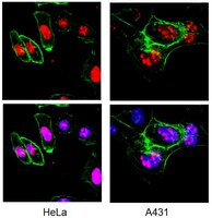


Anti-Ki-67 Antibody, clone Ki-S5 is a high quality Mouse Monoclonal Antibody for the detection of Ki-67 & has been validated in FC, ICC, IHC, IHC(P) & WB.
More>> Anti-Ki-67 Antibody, clone Ki-S5 is a high quality Mouse Monoclonal Antibody for the detection of Ki-67 & has been validated in FC, ICC, IHC, IHC(P) & WB. Less<<Empfohlene Produkte
Übersicht
| Replacement Information |
|---|
| References |
|---|
| Product Information | |
|---|---|
| Format | Purified |
| Control |
|
| Presentation | Mouse monoclonal IgG1 in buffer containing 0.02 M Phosphate buffer, 0.25 M NaCl, pH 7.6 with 0.1% sodium azide. |
| Quality Segment | MQ100 |
| Physicochemical Information |
|---|
| Dimensions |
|---|
| Materials Information |
|---|
| Toxicological Information |
|---|
| Safety Information according to GHS |
|---|
| Safety Information |
|---|
| Storage and Shipping Information | |
|---|---|
| Storage Conditions | Stable for 1 year at 2-8ºC from date of receipt. |
| Packaging Information | |
|---|---|
| Material Size | 25 μg |
| Transport Information |
|---|
| Supplemental Information |
|---|
| Specifications |
|---|
| Global Trade ITEM Number | |
|---|---|
| Bestellnummer | GTIN |
| MAB4190-25UG | 04054839342707 |
Documentation
Anti-Ki-67, clone Ki-S5 Analysenzertifikate
| Titel | Chargennummer |
|---|---|
| Anti-Ki-67, clone Ki-S5 - 3275486 | 3275486 |
| Anti-Ki-67, clone Ki-S5 - 3919632 | 3919632 |
| Anti-Ki-67, clone Ki-S5 - 3990643 | 3990643 |
| Anti-Ki-67, clone Ki-S5 - 4059364 | 4059364 |
| Anti-Ki-67, clone Ki-S5 - 4141174 | 4141174 |
| Anti-Ki-67, clone Ki-S5 - 4315947 | 4315947 |
| Anti-Ki-67, clone Ki-S5 - 4344287 | 4344287 |
Verwandte Produkte & Anwendungen
Produktfamilien
Small Pack Size AntibodiesFind smaller, more convenient sizes of our highly characterized, extensively published and high-quality antibodies.Weitere Informationen >> |