Wenn Sie das Fenster schließen, wird Ihre Konfiguration nicht gespeichert, es sei denn, Sie haben Ihren Artikel in die Bestellung aufgenommen oder zu Ihren Favoriten hinzugefügt.
Klicken Sie auf OK, um das MILLIPLEX® MAP-Tool zu schließen oder auf Abbrechen, um zu Ihrer Auswahl zurückzukehren.
Wählen Sie konfigurierbare Panels & Premixed-Kits - ODER - Kits für die zelluläre Signaltransduktion & MAPmates™
Konfigurieren Sie Ihre MILLIPLEX® MAP-Kits und lassen sich den Preis anzeigen.
Konfigurierbare Panels & Premixed-Kits
Unser breites Angebot enthält Multiplex-Panels, für die Sie die Analyten auswählen können, die am besten für Ihre Anwendung geeignet sind. Unter einem separaten Register können Sie das Premixed-Cytokin-Format oder ein Singleplex-Kit wählen.
Kits für die zelluläre Signaltransduktion & MAPmates™
Wählen Sie gebrauchsfertige Kits zur Erforschung gesamter Signalwege oder Prozesse. Oder konfigurieren Sie Ihre eigenen Kits mit Singleplex MAPmates™.
Die folgenden MAPmates™ sollten nicht zusammen analysiert werden: -MAPmates™, die einen unterschiedlichen Assaypuffer erfordern. -Phosphospezifische und MAPmate™ Gesamtkombinationen wie Gesamt-GSK3β und Gesamt-GSK3β (Ser 9). -PanTyr und locusspezifische MAPmates™, z.B. Phospho-EGF-Rezeptor und Phospho-STAT1 (Tyr701). -Mehr als 1 Phospho-MAPmate™ für ein einziges Target (Akt, STAT3). -GAPDH und β-Tubulin können nicht mit Kits oder MAPmates™, die panTyr enthalten, analysiert werden.
.
Bestellnummer
Bestellinformationen
St./Pkg.
Liste
Dieser Artikel wurde zu Ihren Favoriten hinzugefügt.
Wählen Sie bitte Spezies, Panelart, Kit oder Probenart
Um Ihr MILLIPLEX® MAP-Kit zu konfigurieren, wählen Sie zunächst eine Spezies, eine Panelart und/oder ein Kit.
Custom Premix Selecting "Custom Premix" option means that all of the beads you have chosen will be premixed in manufacturing before the kit is sent to you.
Catalogue Number
Ordering Description
Qty/Pack
List
Dieser Artikel wurde zu Ihren Favoriten hinzugefügt.
Spezies
Panelart
Gewähltes Kit
Menge
Bestellnummer
Bestellinformationen
St./Pkg.
Listenpreis
96-Well Plate
Menge
Bestellnummer
Bestellinformationen
St./Pkg.
Listenpreis
Weitere Reagenzien hinzufügen (MAPmates erfordern die Verwendung eines Puffer- und Detektionskits)
Menge
Bestellnummer
Bestellinformationen
St./Pkg.
Listenpreis
48-602MAG
Buffer Detection Kit for Magnetic Beads
1 Kit
Platzsparende Option Kunden, die mehrere Kits kaufen, können ihre Multiplex-Assaykomponenten in Kunststoffbeuteln anstelle von Packungen erhalten, um eine kompaktere Lagerung zu ermöglichen.
Dieser Artikel wurde zu Ihren Favoriten hinzugefügt.
Das Produkt wurde in Ihre Bestellung aufgenommen
Sie können nun ein weiteres Kit konfigurieren, ein Premixed-Kit wählen, zur Kasse gehen oder das Bestell-Tool schließen.
Anti-dimethyl MBP (Arg107) Antibody is an antibody against dimethyl MBP (Arg107) for use in WB.
More>>Anti-dimethyl MBP (Arg107) Antibody is an antibody against dimethyl MBP (Arg107) for use in WB. Less<<
Anti-dimethyl MBP (Arg107) Antibody: SDB (Sicherheitsdatenblätter), Analysenzertifikate und Qualitätszertifikate, Dossiers, Broschüren und andere verfügbare Dokumente.
Myelin basic protein (MBP) is the major extrinsic membrane protein of central nervous system. MBP is believed to be important in the process of myelination of nerves in the central nervous system (CNS). MBP was initially sequenced in 1979 after isolation from myelin membranes. Since that time, knockout mice deficient in MBP have been developed which showed decreased amounts of CNS myelination and a progressive disorder characterized by tremors, seizures, and early death. The gene for MBP is on chromosome 18; the protein localizes to the CNS and to various cells of the hematopoietic system. The pool of MBP in the central nervous system is very diverse. Not only is MBP present in a number of splice varients, but the protein also undergoes a variety of post-translational modifications, and exists in the CNS in a number of forms (i.e. phosphorylated, methylated, deamidated and citrullinated). Interest in MBP has centered on its role in demyelinating diseases, particularly multiple sclerosis (MS). Several studies have shown a role for antibodies against MBP in the pathogenesis of MS. Some studies have linked a genetic predisposition to MS to the MBP gene, though a majority have not.
References
Product Information
Format
Serum
Control
mouse MBP knockout and wildtype thymus lysates
Presentation
Unpurified rabbit polyclonal in serum containing 0.05% sodium azide.
Applications
Application
Anti-dimethyl MBP (Arg107) Antibody is an antibody against dimethyl MBP (Arg107) for use in WB.
Key Applications
Western Blotting
Biological Information
Immunogen
KLH-conjugated linear peptide corresponding to the C-terminus of MBP dimethylated at Arg107.
Epitope
MBP dimethylated at Arg107
Host
Rabbit
Specificity
This antibody recognizes MBP dimethylated at Arg107.
Isotype
IgG
Species Reactivity
Mouse
Human
Rat
Species Reactivity Note
Demonstrated to react with mouse. Predicted to react with human and rat based on 100% sequence homology.
The protein encoded by the classic MBP gene is a major constituent of the myelin sheath of oligodendrocytes and Schwann cells in the nervous system. However, MBP-related transcripts are also present in the bone marrow and the immune system. These mRNAs arise from the long MBP gene (otherwise called 'Golli-MBP') that contains 3 additional exons located upstream of the classic MBP exons. Alternative splicing from the Golli and the MBP transcription start sites gives rise to 2 sets of MBP-related transcripts and gene products. The Golli mRNAs contain 3 exons unique to Golli-MBP, spliced in-frame to 1 or more MBP exons. They encode hybrid proteins that have N-terminal Golli aa sequence linked to MBP aa sequence. The second family of transcripts contain only MBP exons and produce the well characterized myelin basic proteins. This complex gene structure is conserved among species suggesting that the MBP transcription unit is an integral part of the Golli transcription unit and that this arrangement is important for the function and/or regulation of these genes. [provided by RefSeq].
FUNCTION: The classic group of MBP isoforms (isoform 4-isoform 14) are with PLP the most abundant protein components of the myelin membrane in the CNS. They have a role in both its formation and stabilization. The smaller isoforms might have an important role in remyelination of denuded axons in multiple sclerosis. The non- classic group of MBP isoforms (isoform 1-isoform 3/Golli-MBPs) may preferentially have a role in the early developing brain long before myelination, maybe as components of transcriptional complexes, and may also be involved in signaling pathways in T- cells and neural cells. Differential splicing events combined to optional post-translational modifications give a wide spectrum of isomers, each of them having maybe a specialized function. Induces T-cell proliferation.
SIZE: 304 amino acids; 33117 Da
SUBUNIT: Homodimer; isoform 3 exists as a homodimer.
SUBCELLULAR LOCATION: Myelin membrane; Peripheral membrane protein; Cytoplasmic side. Note=Cytoplasmic side of myelin.
TISSUE SPECIFICITY: MBP isoforms are found in both the central and the peripheral nervous system, whereas Golli-MBP isoforms are expressed in fetal thymus, spleen and spinal cord, as well as in cell lines derived from the immune system.DEVELOPMENTAL STAGE: Expression turns on abruptly in fetus of 14 to 16 weeks. Even smaller isoforms seem to be produced during embryogenesis, some of these persisting in the adult. Expression of isoform MBP2 is more evident at 16 weeks and its relative proportion declined thereafter.
PTM: Several charge isomers of MBP; C1 (the most cationic, least modified, and most abundant form), C2, C3, C4, C5, C6, C7, C8-A and C8-B (the least cationic form); are produced as a result of optional PTM, such as phosphorylation, deamidation of glutamine or asparagine, arginine citrullination and methylation. C8-A and C8-B contain each two mass isoforms termed C8-A(H), C8-A(L), C8-B(H) and C8-B(L), (H) standing for higher and (L) for lower molecular weight. C3, C4 and C5 are phosphorylated. The ratio of methylated arginine residues decreases in aging, making the protein more cationic. & The N-terminal alanine is acetylated (isoform 3, isoform 4, isoform 5 and isoform 6). & Arg-241 was found to be 6% monomethylated and 60% symmetrically dimethylated.
DISEASE: The reduction in the surface charge of citrullinated and/or methylated MBP could result in a weakened attachment to the myelin membrane. This mechanism could be operative in demyelinating diseases such as chronical multiple sclerosis (MS), and fulminating MS (Marburg disease).
SIMILARITY: Belongs to the myelin basic protein family.
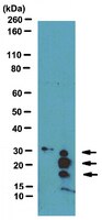
Molecular Weight
Calculated at 33 kDa, the molecular weight of MBP will vary according to specific splice isoforms and modification states. Dimethylated MBP is observed here at 17, 20 and 28 kDa.
Physicochemical Information
Dimensions
Materials Information
Toxicological Information
Safety Information according to GHS
Safety Information
Product Usage Statements
Quality Assurance
Evaluated by Western Blot in mouse MBP knockout and wildtype thymus lysates.
Western Blot Analysis: A 1:1,000 dilution of this antibody detected dimethylated MBP in 10 µg of thymus lysate from a wildtype mouse , while failing to detect dimethylated MBP in a thymus lysate from an MBP knockout.
Usage Statement
Unless otherwise stated in our catalog or other company documentation accompanying the product(s), our products are intended for research use only and are not to be used for any other purpose, which includes but is not limited to, unauthorized commercial uses, in vitro diagnostic uses, ex vivo or in vivo therapeutic uses or any type of consumption or application to humans or animals.
Storage and Shipping Information
Storage Conditions
Stable for 1 year at -20°C from date of receipt. Handling Recommendations: Upon receipt and prior to removing the cap, centrifuge the vial and gently mix the solution. Aliquot into microcentrifuge tubes and store at -20°C. Avoid repeated freeze/thaw cycles, which may damage IgG and affect product performance.
Packaging Information
Material Size
100 µL
Transport Information
Supplemental Information
Specifications
Global Trade ITEM Number
Bestellnummer
GTIN
09-849
04053252288173
Documentation
Literatur
Übersicht
Pub Med ID
Gait abnormalities and progressive myelin degeneration in a new murine model of Pelizaeus-Merzbacher disease with tandem genomic duplication. Clark, K; Sakowski, L; Sperle, K; Banser, L; Landel, CP; Bessert, DA; Skoff, RP; Hobson, GM The Journal of neuroscience : the official journal of the Society for Neuroscience
33
11788-99
2013
Pelizaeus-Merzbacher disease (PMD) is a hypomyelinating leukodystrophy caused by mutations of the proteolipid protein 1 gene (PLP1), which is located on the X chromosome and encodes the most abundant protein of myelin in the central nervous sytem. Approximately 60% of PMD cases result from genomic duplications of a region of the X chromosome that includes the entire PLP1 gene. The duplications are typically in a head-to-tail arrangement, and they vary in size and gene content. Although rodent models with extra copies of Plp1 have been developed, none contains an actual genomic rearrangement that resembles those found in PMD patients. We used mutagenic insertion chromosome engineering resources to generate the Plp1dup mouse model by introducing an X chromosome duplication in the mouse genome that contains Plp1 and five neighboring genes that are also commonly duplicated in PMD patients. The Plp1dup mice display progressive gait abnormalities compared with wild-type littermates. The single duplication leads to increased transcript levels of Plp1 and four of the five other duplicated genes over wild-type levels in the brain beginning the second postnatal week. The Plp1dup mice also display altered transcript levels of other important myelin proteins leading to a progressive degeneration of myelin. Our results show that a single duplication of the Plp1 gene leads to a phenotype similar to the pattern seen in human PMD patients with duplications.
Millipore’s MBP antibodies demonstrate specificity against Myelin basic protein (MBP). See below for related products for MBP, based on the expertise of Upstate & Chemicon. Weitere Informationen >>
Glial Cell Markers
Glial cells, sometimes called neuroglia are non-neuronal cells that maintain homeostasis, form myelin, and provide support and protection for neurons in the brain and peripheral nervous system. Weitere Informationen >>