Wenn Sie das Fenster schließen, wird Ihre Konfiguration nicht gespeichert, es sei denn, Sie haben Ihren Artikel in die Bestellung aufgenommen oder zu Ihren Favoriten hinzugefügt.
Klicken Sie auf OK, um das MILLIPLEX® MAP-Tool zu schließen oder auf Abbrechen, um zu Ihrer Auswahl zurückzukehren.
Wählen Sie konfigurierbare Panels & Premixed-Kits - ODER - Kits für die zelluläre Signaltransduktion & MAPmates™
Konfigurieren Sie Ihre MILLIPLEX® MAP-Kits und lassen sich den Preis anzeigen.
Konfigurierbare Panels & Premixed-Kits
Unser breites Angebot enthält Multiplex-Panels, für die Sie die Analyten auswählen können, die am besten für Ihre Anwendung geeignet sind. Unter einem separaten Register können Sie das Premixed-Cytokin-Format oder ein Singleplex-Kit wählen.
Kits für die zelluläre Signaltransduktion & MAPmates™
Wählen Sie gebrauchsfertige Kits zur Erforschung gesamter Signalwege oder Prozesse. Oder konfigurieren Sie Ihre eigenen Kits mit Singleplex MAPmates™.
Die folgenden MAPmates™ sollten nicht zusammen analysiert werden: -MAPmates™, die einen unterschiedlichen Assaypuffer erfordern. -Phosphospezifische und MAPmate™ Gesamtkombinationen wie Gesamt-GSK3β und Gesamt-GSK3β (Ser 9). -PanTyr und locusspezifische MAPmates™, z.B. Phospho-EGF-Rezeptor und Phospho-STAT1 (Tyr701). -Mehr als 1 Phospho-MAPmate™ für ein einziges Target (Akt, STAT3). -GAPDH und β-Tubulin können nicht mit Kits oder MAPmates™, die panTyr enthalten, analysiert werden.
.
Bestellnummer
Bestellinformationen
St./Pkg.
Liste
Dieser Artikel wurde zu Ihren Favoriten hinzugefügt.
Wählen Sie bitte Spezies, Panelart, Kit oder Probenart
Um Ihr MILLIPLEX® MAP-Kit zu konfigurieren, wählen Sie zunächst eine Spezies, eine Panelart und/oder ein Kit.
Custom Premix Selecting "Custom Premix" option means that all of the beads you have chosen will be premixed in manufacturing before the kit is sent to you.
Catalogue Number
Ordering Description
Qty/Pack
List
Dieser Artikel wurde zu Ihren Favoriten hinzugefügt.
Spezies
Panelart
Gewähltes Kit
Menge
Bestellnummer
Bestellinformationen
St./Pkg.
Listenpreis
96-Well Plate
Menge
Bestellnummer
Bestellinformationen
St./Pkg.
Listenpreis
Weitere Reagenzien hinzufügen (MAPmates erfordern die Verwendung eines Puffer- und Detektionskits)
Menge
Bestellnummer
Bestellinformationen
St./Pkg.
Listenpreis
48-602MAG
Buffer Detection Kit for Magnetic Beads
1 Kit
Platzsparende Option Kunden, die mehrere Kits kaufen, können ihre Multiplex-Assaykomponenten in Kunststoffbeuteln anstelle von Packungen erhalten, um eine kompaktere Lagerung zu ermöglichen.
Dieser Artikel wurde zu Ihren Favoriten hinzugefügt.
Das Produkt wurde in Ihre Bestellung aufgenommen
Sie können nun ein weiteres Kit konfigurieren, ein Premixed-Kit wählen, zur Kasse gehen oder das Bestell-Tool schließen.
Anti-ABCA4, clone 5B4, Cat. No. MABN2440, is a highly specific mouse monoclonal antibody that targets Retinal-specific ATP-binding cassette transporter and has been tested for use in Immunocytochemistry, Immunoprecipitation, and Western Blotting.
More>>Anti-ABCA4, clone 5B4, Cat. No. MABN2440, is a highly specific mouse monoclonal antibody that targets Retinal-specific ATP-binding cassette transporter and has been tested for use in Immunocytochemistry, Immunoprecipitation, and Western Blotting. Less<<
Anti-ABCA4 Antibody, clone 5B4: SDB (Sicherheitsdatenblätter), Analysenzertifikate und Qualitätszertifikate, Dossiers, Broschüren und andere verfügbare Dokumente.
Retinal-specific ATP-binding cassette transporter (UniProt: P78363; also known as ATP-binding cassette sub-family A member 4, ABCA4, RIM ABC transporter, RIM protein, RmP, Stargardt disease protein) is encoded by the ABCA4 (also known as ABCR) gene (Gene ID: 24) in human. ABCA4 is a multi-pass membrane protein that is exclusively expressed in retina and is found in the rims of rod photoreceptor cells. It contains two ABC transporter domains (aa 929-1160 and 1938-2170). In the visual cycle, it act as a retinal extruder or retinal-phosphatidylethanolamine flippase to facilitate the removal of all-trans-retinal from disc membranes following the photobleaching of rhodopsin. Mutations in ABCA4 gene are reported to cause Stargardt disease, which is characterized by decreased central vision, atrophy of the macula and underlying retinal pigment epithelium, and frequent presence of prominent flecks in the posterior pole of the retina. It has also been linked to age-related macular degeneration and retinitis pigmentosa.
References
Product Information
Format
Purified
Presentation
Purified mouse monoclonal antibody IgG2b in buffer containing 0.1 M Tris-Glycine (pH 7.4), 150 mM NaCl with 0.05% sodium azide.
Anti-ABCA4, clone 5B4, Cat. No. MABN2440, is a highly specific mouse monoclonal antibody that targets Retinal-specific ATP-binding cassette transporter and has been tested for use in Immunocytochemistry, Immunoprecipitation, and Western Blotting.
Key Applications
Immunocytochemistry
Western Blotting
Immunoprecipitation
Application Notes
Immunocytochemistry Analysis: A representative lot detected ABCA4 in Immunocytochemistry applications (Ahn, J., et. al. (2003). J Biol Chem. 278(41):39600-8).
Western Blotting Analysis: A representative lot detected ABCA4 in Western Blotting applications (Ahn, J., et. al. (2003). J Biol Chem. 278(41):39600-8; Bungert, S., et. al. (2001). J Biol Chem. 276(26):23539-46).
Immunoprecipitation Analysis: A representative lot detected ABCA4 in Immunoprecipitation applications (Ahn, J., et. al. (2003). J Biol Chem. 278(41):39600-8; Bungert, S., et. al. (2001). J Biol Chem. 276(26):23539-46).
Biological Information
Immunogen
GST-tagged recombinant fragment corresponding to 249 amino acids from the N-terminal half of human Retinal-specific ATP-binding cassette transporter.
Clone
5B4
Concentration
Please refer to lot specific datasheet.
Host
Mouse
Specificity
Clone 5B4 detects Retinal-specific ATP-binding cassette transporter protein in human and bovine. It targets an epitope within 249 amino acids from the N-terminal half.
Isotype
IgG2bκ
Species Reactivity
Bovine
Human
Species Reactivity Note
Human, Bovine. Predicted to react with Mouse based on 100% sequence homology.
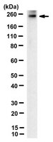
250 kDa observed; 255.94 kDa calculated. Uncharacterized bands may be observed in some lysate(s).
Physicochemical Information
Dimensions
Materials Information
Toxicological Information
Safety Information according to GHS
Safety Information
Product Usage Statements
Quality Assurance
Evaluated by Western Blotting in human retina tissue lysate.
Western Blotting Analysis: 1 µg/mL of this antibody detected ABCA4 in 10 µg of human retina tissue lysate.
Usage Statement
Unless otherwise stated in our catalog or other company documentation accompanying the product(s), our products are intended for research use only and are not to be used for any other purpose, which includes but is not limited to, unauthorized commercial uses, in vitro diagnostic uses, ex vivo or in vivo therapeutic uses or any type of consumption or application to humans or animals.