Wenn Sie das Fenster schließen, wird Ihre Konfiguration nicht gespeichert, es sei denn, Sie haben Ihren Artikel in die Bestellung aufgenommen oder zu Ihren Favoriten hinzugefügt.
Klicken Sie auf OK, um das MILLIPLEX® MAP-Tool zu schließen oder auf Abbrechen, um zu Ihrer Auswahl zurückzukehren.
Wählen Sie konfigurierbare Panels & Premixed-Kits - ODER - Kits für die zelluläre Signaltransduktion & MAPmates™
Konfigurieren Sie Ihre MILLIPLEX® MAP-Kits und lassen sich den Preis anzeigen.
Konfigurierbare Panels & Premixed-Kits
Unser breites Angebot enthält Multiplex-Panels, für die Sie die Analyten auswählen können, die am besten für Ihre Anwendung geeignet sind. Unter einem separaten Register können Sie das Premixed-Cytokin-Format oder ein Singleplex-Kit wählen.
Kits für die zelluläre Signaltransduktion & MAPmates™
Wählen Sie gebrauchsfertige Kits zur Erforschung gesamter Signalwege oder Prozesse. Oder konfigurieren Sie Ihre eigenen Kits mit Singleplex MAPmates™.
Die folgenden MAPmates™ sollten nicht zusammen analysiert werden: -MAPmates™, die einen unterschiedlichen Assaypuffer erfordern. -Phosphospezifische und MAPmate™ Gesamtkombinationen wie Gesamt-GSK3β und Gesamt-GSK3β (Ser 9). -PanTyr und locusspezifische MAPmates™, z.B. Phospho-EGF-Rezeptor und Phospho-STAT1 (Tyr701). -Mehr als 1 Phospho-MAPmate™ für ein einziges Target (Akt, STAT3). -GAPDH und β-Tubulin können nicht mit Kits oder MAPmates™, die panTyr enthalten, analysiert werden.
.
Bestellnummer
Bestellinformationen
St./Pkg.
Liste
Dieser Artikel wurde zu Ihren Favoriten hinzugefügt.
Wählen Sie bitte Spezies, Panelart, Kit oder Probenart
Um Ihr MILLIPLEX® MAP-Kit zu konfigurieren, wählen Sie zunächst eine Spezies, eine Panelart und/oder ein Kit.
Custom Premix Selecting "Custom Premix" option means that all of the beads you have chosen will be premixed in manufacturing before the kit is sent to you.
Catalogue Number
Ordering Description
Qty/Pack
List
Dieser Artikel wurde zu Ihren Favoriten hinzugefügt.
Spezies
Panelart
Gewähltes Kit
Menge
Bestellnummer
Bestellinformationen
St./Pkg.
Listenpreis
96-Well Plate
Menge
Bestellnummer
Bestellinformationen
St./Pkg.
Listenpreis
Weitere Reagenzien hinzufügen (MAPmates erfordern die Verwendung eines Puffer- und Detektionskits)
Menge
Bestellnummer
Bestellinformationen
St./Pkg.
Listenpreis
48-602MAG
Buffer Detection Kit for Magnetic Beads
1 Kit
Platzsparende Option Kunden, die mehrere Kits kaufen, können ihre Multiplex-Assaykomponenten in Kunststoffbeuteln anstelle von Packungen erhalten, um eine kompaktere Lagerung zu ermöglichen.
Dieser Artikel wurde zu Ihren Favoriten hinzugefügt.
Das Produkt wurde in Ihre Bestellung aufgenommen
Sie können nun ein weiteres Kit konfigurieren, ein Premixed-Kit wählen, zur Kasse gehen oder das Bestell-Tool schließen.
MABT1240
Sigma-AldrichAnti-Cathepsin G Antibody, clone AHN-11, Ascites Free
This Anti-Cathepsin G Antibody, clone AHN-11, Ascites Free is validated for use in Immunocytochemistry, Western Blotting, ELISA, Radioimmunoassay for the detection of Cathepsin G.
More>>This Anti-Cathepsin G Antibody, clone AHN-11, Ascites Free is validated for use in Immunocytochemistry, Western Blotting, ELISA, Radioimmunoassay for the detection of Cathepsin G. Less<<
SDB (Sicherheitsdatenblätter), Analysenzertifikate und Qualitätszertifikate, Dossiers, Broschüren und andere verfügbare Dokumente.
Anti-Cathepsin G Antibody, clone AHN-11, Ascites Free
Alternate Names
Cathepsin G
CatG
CG
Background Information
Cathepsin G (EC 3.4.21.20; UniProt P08311; also known as CatG, CG) is encoded by the CTSG gene (Gene ID 1511) in human. Cathepsin G is a peptidase S1 protein family protease found in azurophil granules of neutrophilic polymorphonuclear leukocytes. Cathepsin G displays a substrate specificity similar to that of chymotrypsin C, but it is most closely related to other immune serine proteases, such as neutrophil elastase and the granzymes. Cathepsin G is involved in connective tissue remodeling at sites of inflammation. Cathepsin G is also a component of neutrophil extracellular traps (NETs) composed of ejected lattice of chromatin enmeshed with granular and nuclear proteins, including histones, calprotectin and cathepsin G, that are capable of capturing and killing microbial invaders. Antibodies raised against cathepsin G or histones have been shown to limit the bactericidal capacity of NETs towards pathogens. Cathepsin G is prouduced with a signal peptide (a.a. 1-18) and a propeptide sequence (a.a. 19-20) that are removed posttranslationally to yield the mature (a.a. 21-255) enzyme.
References
Product Information
Format
Purified
Presentation
Purified mouse monoclonal IgG2aκ antibody in buffer containing 0.1 M Tris-Glycine (pH 7.4), 150 mM NaCl with 0.05% sodium azide.
This Anti-Cathepsin G Antibody, clone AHN-11, Ascites Free is validated for use in Immunocytochemistry, Western Blotting, ELISA, Radioimmunoassay for the detection of Cathepsin G.
Key Applications
Immunocytochemistry
Western Blotting
ELISA
Radioimmunoassay
Application Notes
Western Blotting Analysis: A representative lot detected cathepsin G in the primary granule fraction from human neutrophils (Stroncek D.F., et al. (2005). J. Transl. Med. 3:36). Immunocytochemistry Analysis: Clone AHN-11 hybridoma culture supernatant immunostained ethanol-fixed human neutrophils, but not other peripheral blood cells, including platelets, lymphocytes, and eosinophils. Faint immunostaining of some monocytes was occasionally detected (Skubitz, K.M., et al. (1989). J. Leukoc. Biol. 46(2):109-118). ELISA Analysis: A representative lot captured cathepsin G from human neutrophil granule extract. The captured cathepsin G served to detect the presence of serum cathepsin G autoantibody levels in patients serum samples (Ellerbroek, P.M., et al. (1994). J. Clin. Pathol. 47(3):257-262). Radioimmunoassay Analysis: Clone AHN-11 hybridoma culture supernatant bound to well surface coated with dried human neutrophils, but not platelets or erythrocytes by solid-phase radioimmunoassay (Skubitz, K.M., et al. (1989). J. Leukoc. Biol. 46(2):109-118). Radioimmunoassay Analysis: Clone AHN-11 hybridoma culture supernatant bound to well surface coated with content of primary granule, but not secondary granule, plasma membrane, or cytoplasm fraction from neutrophils by solid-phase radioimmunoassay (Skubitz, K.M., et al. (1989). J. Leukoc. Biol. 46(2):109-118). Radioimmunoassay Analysis: Clone AHN-11 hybridoma culture supernatant reacted with purified human cathepsin G, but not elastase, EPO, esterase N, proteinase 3, plamin, kallidrein, lactoferrin, MBP, or thrombin by solid-phase radioimmunoassay (Skubitz, K.M., et al. (1989). J. Leukoc. Biol. 46(2):109-118).
Biological Information
Immunogen
Human neutrophils.
Clone
AHN-11
Concentration
Please refer to lot specific datasheet.
Host
Mouse
Specificity
Clone AHN-11 reacted specifically with neutrophil cathepsin G, but not the homologous neutrophil neutral proteases, elastase, proteinase 3, or esterase N (Skubitz, K.M., et al. (1989). J. Leukoc. Biol. 46(2):109-118).
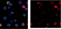
Immunocytochemistry Analysis: A 1:200 dilution of this antibody detected Cathepsin G in HL60 human promyelocytic leukemia cells.
Usage Statement
Unless otherwise stated in our catalog or other company documentation accompanying the product(s), our products are intended for research use only and are not to be used for any other purpose, which includes but is not limited to, unauthorized commercial uses, in vitro diagnostic uses, ex vivo or in vivo therapeutic uses or any type of consumption or application to humans or animals.
Storage and Shipping Information
Storage Conditions
Stable for 1 year at 2-8°C from date of receipt.
Packaging Information
Material Size
100 μg
Transport Information
Supplemental Information
Specifications
Global Trade ITEM Number
Bestellnummer
GTIN
MABT1240
04055977353600
Documentation
Anti-Cathepsin G Antibody, clone AHN-11, Ascites Free SDB