Wenn Sie das Fenster schließen, wird Ihre Konfiguration nicht gespeichert, es sei denn, Sie haben Ihren Artikel in die Bestellung aufgenommen oder zu Ihren Favoriten hinzugefügt.
Klicken Sie auf OK, um das MILLIPLEX® MAP-Tool zu schließen oder auf Abbrechen, um zu Ihrer Auswahl zurückzukehren.
Wählen Sie konfigurierbare Panels & Premixed-Kits - ODER - Kits für die zelluläre Signaltransduktion & MAPmates™
Konfigurieren Sie Ihre MILLIPLEX® MAP-Kits und lassen sich den Preis anzeigen.
Konfigurierbare Panels & Premixed-Kits
Unser breites Angebot enthält Multiplex-Panels, für die Sie die Analyten auswählen können, die am besten für Ihre Anwendung geeignet sind. Unter einem separaten Register können Sie das Premixed-Cytokin-Format oder ein Singleplex-Kit wählen.
Kits für die zelluläre Signaltransduktion & MAPmates™
Wählen Sie gebrauchsfertige Kits zur Erforschung gesamter Signalwege oder Prozesse. Oder konfigurieren Sie Ihre eigenen Kits mit Singleplex MAPmates™.
Die folgenden MAPmates™ sollten nicht zusammen analysiert werden: -MAPmates™, die einen unterschiedlichen Assaypuffer erfordern. -Phosphospezifische und MAPmate™ Gesamtkombinationen wie Gesamt-GSK3β und Gesamt-GSK3β (Ser 9). -PanTyr und locusspezifische MAPmates™, z.B. Phospho-EGF-Rezeptor und Phospho-STAT1 (Tyr701). -Mehr als 1 Phospho-MAPmate™ für ein einziges Target (Akt, STAT3). -GAPDH und β-Tubulin können nicht mit Kits oder MAPmates™, die panTyr enthalten, analysiert werden.
.
Bestellnummer
Bestellinformationen
St./Pkg.
Liste
Dieser Artikel wurde zu Ihren Favoriten hinzugefügt.
Wählen Sie bitte Spezies, Panelart, Kit oder Probenart
Um Ihr MILLIPLEX® MAP-Kit zu konfigurieren, wählen Sie zunächst eine Spezies, eine Panelart und/oder ein Kit.
Custom Premix Selecting "Custom Premix" option means that all of the beads you have chosen will be premixed in manufacturing before the kit is sent to you.
Catalogue Number
Ordering Description
Qty/Pack
List
Dieser Artikel wurde zu Ihren Favoriten hinzugefügt.
Spezies
Panelart
Gewähltes Kit
Menge
Bestellnummer
Bestellinformationen
St./Pkg.
Listenpreis
96-Well Plate
Menge
Bestellnummer
Bestellinformationen
St./Pkg.
Listenpreis
Weitere Reagenzien hinzufügen (MAPmates erfordern die Verwendung eines Puffer- und Detektionskits)
Menge
Bestellnummer
Bestellinformationen
St./Pkg.
Listenpreis
48-602MAG
Buffer Detection Kit for Magnetic Beads
1 Kit
Platzsparende Option Kunden, die mehrere Kits kaufen, können ihre Multiplex-Assaykomponenten in Kunststoffbeuteln anstelle von Packungen erhalten, um eine kompaktere Lagerung zu ermöglichen.
Dieser Artikel wurde zu Ihren Favoriten hinzugefügt.
Das Produkt wurde in Ihre Bestellung aufgenommen
Sie können nun ein weiteres Kit konfigurieren, ein Premixed-Kit wählen, zur Kasse gehen oder das Bestell-Tool schließen.
ABS983
Sigma-AldrichAnti-DMT1 Antibody
This Anti-DMT1 Antibody is validated for use in Western Blotting and Immunocytochemistry for the detection of DMT1.
More>>This Anti-DMT1 Antibody is validated for use in Western Blotting and Immunocytochemistry for the detection of DMT1. Less<<
Anti-DMT1 Antibody: SDB (Sicherheitsdatenblätter), Analysenzertifikate und Qualitätszertifikate, Dossiers, Broschüren und andere verfügbare Dokumente.
Divalent metal ion transporter 1 (DMT1) is a proton-coupled Fe2+ transporter that is essential for iron uptake in enterocytes and for transferrin-associated endosomal iron transport in many other cell types. DMT1 dysfunction is associated with several diseases such as iron overload disorders and neurodegenerative diseases. DMT1 can transport manganese, cobalt, cadmium, nickel, and vanadium. DMT1 is also involved in iron transport from acidified endosomes into the cytoplasm in erythroid precursor cells as well as liver internal metabolism of iron. Interestingly too, in mammalian cells, mitochondria receive most incoming iron, and the outer mitochondrial membrane appears to be a major location for DMT1, which appears to be the major importer of iron into the mitochondria.
This Anti-DMT1 Antibody is validated for use in Western Blotting and Immunocytochemistry for the detection of DMT1.
Key Applications
Western Blotting
Immunocytochemistry
Application Notes
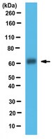
Western Blot Analysis: 2 µg/ml of this antibody detected DMT1 in 50 µg of mouse liver, spleen and duodenum tissue lysate (Schumann, K.,et al. (2010). Journal of Trace Elements in Medicine and Biology 24 (2010) 58–66) Immunocytochemistry Analysis: A 1:100 dilution of this antibody from a representative lot detected DMT1 in Hela, A431, and N1H-3T3 cells
Evaluated by Western Blotting in human placenta tissue lysate.
Western Blotting Analysis: A 1:1,000 dilution of this antibody detected DMT1 in 10 µg of human placenta tissue lysate.
Usage Statement
Unless otherwise stated in our catalog or other company documentation accompanying the product(s), our products are intended for research use only and are not to be used for any other purpose, which includes but is not limited to, unauthorized commercial uses, in vitro diagnostic uses, ex vivo or in vivo therapeutic uses or any type of consumption or application to humans or animals.
Storage and Shipping Information
Storage Conditions
Stable for 1 year at -20°C from date of receipt. Handling Recommendations: Upon receipt and prior to removing the cap, centrifuge the vial and gently mix the solution. Aliquot into microcentrifuge tubes and store at -20°C. Avoid repeated freeze/thaw cycles, which may damage IgG and affect product performance.
Iron absorption and distribution in TNF(DeltaARE/+) mice, a model of chronic inflammation. Schümann, Klaus, et al. J Trace Elem Med Biol, 24: 58-66 (2010)
2009
Hemizygous TNF(DeltaARE/+) mice are a murine model for chronic inflammation. We utilized these animals to study iron-kinetics and corresponding protein expression in an iron-deficient and iron-adequate setting. (59)Fe-absorption was determined in ligated duodenal loops in vivo. Whole body distribution of i.v. injected (59)Fe was analysed, and the organ specific expression of ferroportin, transferrin receptor-1, hepcidin and duodenal DMT-1 was quantified by real-time PCR and Western blotting. Duodenal (59)Fe-lumen-to-body transport was not affected by the genotype. Duodenal (59)Fe-retention was increased in TNF(DeltaARE/+) mice, suggesting higher (59)Fe-losses with defoliated enterocytes. Iron-deficiency increased duodenal (59)Fe-lumen-to-body transport, and higher duodenal (59)Fe-tissue retention went along with higher duodenal DMT-1, ferroportin, and liver hepcidin expression. TNF(DeltaARE/+) mice significantly increase their (59)Fe-content in inflamed joints and ilea, and correspondingly reduce splenic (59)Fe-content. Leukocyte infiltrations in the joints suggest a substantial shift of iron-loaded RES cells to inflamed tissues as the underlying mechanism. This finding was paralleled by increased non-haem iron content in joints and reduced haemoglobin and haematocrit concentrations in TNF(DeltaARE/+) mice. In conclusion, erythropoiesis in inflamed TNF(DeltaARE/+) mice could be iron-limited due to losses with exfoliated iron-loaded enterocytes and/or to increased iron-retention in RES cells that shift from the spleen to inflamed tissues.