Wenn Sie das Fenster schließen, wird Ihre Konfiguration nicht gespeichert, es sei denn, Sie haben Ihren Artikel in die Bestellung aufgenommen oder zu Ihren Favoriten hinzugefügt.
Klicken Sie auf OK, um das MILLIPLEX® MAP-Tool zu schließen oder auf Abbrechen, um zu Ihrer Auswahl zurückzukehren.
Wählen Sie konfigurierbare Panels & Premixed-Kits - ODER - Kits für die zelluläre Signaltransduktion & MAPmates™
Konfigurieren Sie Ihre MILLIPLEX® MAP-Kits und lassen sich den Preis anzeigen.
Konfigurierbare Panels & Premixed-Kits
Unser breites Angebot enthält Multiplex-Panels, für die Sie die Analyten auswählen können, die am besten für Ihre Anwendung geeignet sind. Unter einem separaten Register können Sie das Premixed-Cytokin-Format oder ein Singleplex-Kit wählen.
Kits für die zelluläre Signaltransduktion & MAPmates™
Wählen Sie gebrauchsfertige Kits zur Erforschung gesamter Signalwege oder Prozesse. Oder konfigurieren Sie Ihre eigenen Kits mit Singleplex MAPmates™.
Die folgenden MAPmates™ sollten nicht zusammen analysiert werden: -MAPmates™, die einen unterschiedlichen Assaypuffer erfordern. -Phosphospezifische und MAPmate™ Gesamtkombinationen wie Gesamt-GSK3β und Gesamt-GSK3β (Ser 9). -PanTyr und locusspezifische MAPmates™, z.B. Phospho-EGF-Rezeptor und Phospho-STAT1 (Tyr701). -Mehr als 1 Phospho-MAPmate™ für ein einziges Target (Akt, STAT3). -GAPDH und β-Tubulin können nicht mit Kits oder MAPmates™, die panTyr enthalten, analysiert werden.
.
Bestellnummer
Bestellinformationen
St./Pkg.
Liste
Dieser Artikel wurde zu Ihren Favoriten hinzugefügt.
Wählen Sie bitte Spezies, Panelart, Kit oder Probenart
Um Ihr MILLIPLEX® MAP-Kit zu konfigurieren, wählen Sie zunächst eine Spezies, eine Panelart und/oder ein Kit.
Custom Premix Selecting "Custom Premix" option means that all of the beads you have chosen will be premixed in manufacturing before the kit is sent to you.
Catalogue Number
Ordering Description
Qty/Pack
List
Dieser Artikel wurde zu Ihren Favoriten hinzugefügt.
Spezies
Panelart
Gewähltes Kit
Menge
Bestellnummer
Bestellinformationen
St./Pkg.
Listenpreis
96-Well Plate
Menge
Bestellnummer
Bestellinformationen
St./Pkg.
Listenpreis
Weitere Reagenzien hinzufügen (MAPmates erfordern die Verwendung eines Puffer- und Detektionskits)
Menge
Bestellnummer
Bestellinformationen
St./Pkg.
Listenpreis
48-602MAG
Buffer Detection Kit for Magnetic Beads
1 Kit
Platzsparende Option Kunden, die mehrere Kits kaufen, können ihre Multiplex-Assaykomponenten in Kunststoffbeuteln anstelle von Packungen erhalten, um eine kompaktere Lagerung zu ermöglichen.
Dieser Artikel wurde zu Ihren Favoriten hinzugefügt.
Das Produkt wurde in Ihre Bestellung aufgenommen
Sie können nun ein weiteres Kit konfigurieren, ein Premixed-Kit wählen, zur Kasse gehen oder das Bestell-Tool schließen.
Anti-MLKL, clone 5A6, Cat. No. MABC1634, is a rat monoclonal antibody that detects MLKL and is tested for use in Immunofluorescence and Western Blotting.
More>>Anti-MLKL, clone 5A6, Cat. No. MABC1634, is a rat monoclonal antibody that detects MLKL and is tested for use in Immunofluorescence and Western Blotting. Less<<
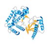
Mixed lineage kinase domain-like protein (UniProt: Q9D2Y4; also known as MLKL) is encoded by the Mlkl gene (Gene ID: 74568) in murine species. MLKL is a pseudokinase that is a terminal protein in the pro-inflammatory necroptotic cell death program. It plays a critical role in TNFa-induced necroptosis. It is highly expressed in thymus, colon, intestine, liver, spleen, and lung. Its expression is much lower in skeletal muscle, heart and kidney, and it is not detected in the brain. Although it contains a protein kinase domain (aa 192-456), it lacks several residues that are essential for protein kinase activity. It is activated following phosphorylation by RIPK3, which leads to its homotrimerization and localization to the plasma membrane where it binds to highly phosphorylated inositol (InsP6) and disrupts membrane integrity to cause necrotic cell death. Phosphorylation of MLKL at serine 345 is shown to be essential for necroptosis in murine cells. Its interaction with RIPK3 is species specific and mouse MLKL does not interact with human RIPK3. In contrast to human protein, mouse MLKL is not inhibited by necrosulfonamide, because at position 85 it contains a tryptophan residue instead of a cysteine. Under basal conditions, MLKL is shown to reside in small puncta that are distributed evenly throughout the cytoplasm, but it is detected in plasma membrane shortly after its phosphorylation. It contains two coiled coil regions (aa 61-81 and 138-229) and the second coiled coil region is responsible for its oligomerization. Two isoforms of MLKL have been described that are produced by alternative splicing. (Ref.: Samson, AL., et al. (2021). Cell Death Differ. 28(7); 2126-2144; Wang, H., et al (2014). Mol. Cell 54(1); 133-146).
References
Product Information
Format
Purified
Presentation
Purified rat monoclonal antibody IgG2a in buffer containing 0.1 M Tris-Glycine (pH 7.4), 150 mM NaCl with 0.05% sodium azide
Anti-MLKL, clone 5A6, Cat. No. MABC1634, is a rat monoclonal antibody that detects MLKL and is tested for use in Immunofluorescence and Western Blotting.
Key Applications
Western Blotting
Immunofluorescence
Application Notes
Tested Applications
Immunofluorescence Analysis: A representative lot detected MLKL in Immunofluorescence applications (Samson, A.L., et al. (2021). Cell Death Differ. 28(7):2126-2144).
Western Blotting Analysis: A representative lot detected MLKL in Western Blotting applications (Samson, A.L., et al. (2021). Cell Death Differ. 28(7):2126-2144).
Note: Actual optimal working dilutions must be determined by end user as specimens, and experimental conditions may vary with the end user
Biological Information
Immunogen
His-tagged full-length recombinant mouse Mixed lineage kinase domain-like protein (MLKL).
Epitope
C-terminal
Clone
5A6
Concentration
0.5 mg/mL. Please refer to guidance on suggested starting dilutions and/or titers per application and sample type.
Host
Rat
Specificity
Clone 5A6 is a rat monoclonal antibody that detects murine Mixed lineage kinase domain-like protein (MLKL). It targets an epitope within the C-terminal region.
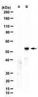
~54 kDa observed; 54.32 kDa calculated. Uncharacterized bands may be observed in some lysate(s).
Physicochemical Information
Dimensions
Materials Information
Toxicological Information
Safety Information according to GHS
Safety Information
Product Usage Statements
Quality Assurance
Evaluated by Western Blotting in lysates from Mouse dermal fibroblasts.
Western Blotting Analysis (WB): A 1:500 dilution of this antibody detected MLKL in lysates from wild-type Mouse dermal fibroblasts, but not in lysates from fibroblasts with Mlkl knockout.
Usage Statement
Unless otherwise stated in our catalog or other company documentation accompanying the product(s), our products are intended for research use only and are not to be used for any other purpose, which includes but is not limited to, unauthorized commercial uses, in vitro diagnostic uses, ex vivo or in vivo therapeutic uses or any type of consumption or application to humans or animals.