Wenn Sie das Fenster schließen, wird Ihre Konfiguration nicht gespeichert, es sei denn, Sie haben Ihren Artikel in die Bestellung aufgenommen oder zu Ihren Favoriten hinzugefügt.
Klicken Sie auf OK, um das MILLIPLEX® MAP-Tool zu schließen oder auf Abbrechen, um zu Ihrer Auswahl zurückzukehren.
Wählen Sie konfigurierbare Panels & Premixed-Kits - ODER - Kits für die zelluläre Signaltransduktion & MAPmates™
Konfigurieren Sie Ihre MILLIPLEX® MAP-Kits und lassen sich den Preis anzeigen.
Konfigurierbare Panels & Premixed-Kits
Unser breites Angebot enthält Multiplex-Panels, für die Sie die Analyten auswählen können, die am besten für Ihre Anwendung geeignet sind. Unter einem separaten Register können Sie das Premixed-Cytokin-Format oder ein Singleplex-Kit wählen.
Kits für die zelluläre Signaltransduktion & MAPmates™
Wählen Sie gebrauchsfertige Kits zur Erforschung gesamter Signalwege oder Prozesse. Oder konfigurieren Sie Ihre eigenen Kits mit Singleplex MAPmates™.
Die folgenden MAPmates™ sollten nicht zusammen analysiert werden: -MAPmates™, die einen unterschiedlichen Assaypuffer erfordern. -Phosphospezifische und MAPmate™ Gesamtkombinationen wie Gesamt-GSK3β und Gesamt-GSK3β (Ser 9). -PanTyr und locusspezifische MAPmates™, z.B. Phospho-EGF-Rezeptor und Phospho-STAT1 (Tyr701). -Mehr als 1 Phospho-MAPmate™ für ein einziges Target (Akt, STAT3). -GAPDH und β-Tubulin können nicht mit Kits oder MAPmates™, die panTyr enthalten, analysiert werden.
.
Bestellnummer
Bestellinformationen
St./Pkg.
Liste
Dieser Artikel wurde zu Ihren Favoriten hinzugefügt.
Wählen Sie bitte Spezies, Panelart, Kit oder Probenart
Um Ihr MILLIPLEX® MAP-Kit zu konfigurieren, wählen Sie zunächst eine Spezies, eine Panelart und/oder ein Kit.
Custom Premix Selecting "Custom Premix" option means that all of the beads you have chosen will be premixed in manufacturing before the kit is sent to you.
Catalogue Number
Ordering Description
Qty/Pack
List
Dieser Artikel wurde zu Ihren Favoriten hinzugefügt.
Spezies
Panelart
Gewähltes Kit
Menge
Bestellnummer
Bestellinformationen
St./Pkg.
Listenpreis
96-Well Plate
Menge
Bestellnummer
Bestellinformationen
St./Pkg.
Listenpreis
Weitere Reagenzien hinzufügen (MAPmates erfordern die Verwendung eines Puffer- und Detektionskits)
Menge
Bestellnummer
Bestellinformationen
St./Pkg.
Listenpreis
48-602MAG
Buffer Detection Kit for Magnetic Beads
1 Kit
Platzsparende Option Kunden, die mehrere Kits kaufen, können ihre Multiplex-Assaykomponenten in Kunststoffbeuteln anstelle von Packungen erhalten, um eine kompaktere Lagerung zu ermöglichen.
Dieser Artikel wurde zu Ihren Favoriten hinzugefügt.
Das Produkt wurde in Ihre Bestellung aufgenommen
Sie können nun ein weiteres Kit konfigurieren, ein Premixed-Kit wählen, zur Kasse gehen oder das Bestell-Tool schließen.
ABC1451-100UG
Sigma-AldrichAnti-PDGF-D
Anti-PDGF-D, Cat. No. ABC1451, is a rabbit polyclonal antibody that detects Platelet-derived growth factor D in human cells and has been tested for use in Western Blotting.
More>>Anti-PDGF-D, Cat. No. ABC1451, is a rabbit polyclonal antibody that detects Platelet-derived growth factor D in human cells and has been tested for use in Western Blotting. Less<<
Anti-PDGF-D: SDB (Sicherheitsdatenblätter), Analysenzertifikate und Qualitätszertifikate, Dossiers, Broschüren und andere verfügbare Dokumente.
Platelet-derived growth factor D (UniProt: Q9GZP0; also known as PDGF-D, Iris-expressed growth factor, Spinal cord-derived growth factor B, SCDGF-B) is encoded by the PDGFD (also known as IEGF, SCDGFB, MSTP036, UNQ1899/PRO4345) gene (Gene ID: 80310) in human. Four PDGF ligands known as PDGF-A, PDGF-B, PDGF-C, and PDGF-D have been described that exert their effect by binding to their cognate receptors. PDGF-D is expressed at high levels in the heart, pancreas, adrenal gland and ovary and at low levels in placenta, liver, kidney, prostate, testis, small intestine, spleen, and colon. PDGF-D is synthesized with a signal peptide (aa 1-18), which is subsequently removed. The latent form of PDGF-D contains a CUB domain at its N-terminal region, a growth factor domain and a hinge region in between. The latent form is activated by the proteolytic action of matriptase that removes the CUB domain, which results in unmasking of the receptor-binding epitopes on the core domain. YR247GR249S within the PDGF-D hinge region is shown to be the cleavage site for removal of the inhibitory CUB domain. PDGF-D plays an important role in wound healing and induces macrophage recruitment, increased interstitial pressure, and blood vessel maturation during angiogenesis. PDGF-D also induces cellular transformation and promotes tumor growth by accelerating the proliferation rate of tumor cells and by stimulation of tumor neovascularization. (Ref.: Huang, W., and Kim, H-R, C (2015). J. Biol. Chem. 290(14); 9162-9170).
References
Product Information
Format
Purified
Presentation
Purified rabbit polyclonal antibody in PBS (pH 7.4) with 0.09% sodium azide.
Applications
Application
Anti-PDGF-D, Cat. No. ABC1451, is a rabbit polyclonal antibody that detects Platelet-derived growth factor D in human cells and has been tested for use in Western Blotting.
Key Applications
Western Blotting
Application Notes
Western Blotting Analysis: A representative lot detected PDGF-D in human recombinant-PDGF D (Huang, W., et. al. (2015). J Biol Chem. 290(14):9162-70) and LNCaP cells expressing PDGF-D, BS-C-1 cells transfected with the plasmid pTF7-PDGF D:HIS, recombinant PDGF-D (Ustach, C.V., et. al. (2004). Cancer Res. 64(5):1722-9).
Western Blotting Analysis: A representative lot detected PDGF-D in PBS exchanged condition media collected from LNCaP cells transfected with PDGF-D expression vector (Courtesy of Hyeong-Reh Choi Kim lab at Wayne State University).
Biological Information
Immunogen
A linear peptide corresponding to 18 amino acids from the C-terminal half of human Platelet-derived growth factor D.
Epitope
unknown
Concentration
Please refer to lot specific datasheet.
Host
Rabbit
Specificity
This rabbit polyclonal antibody detects Platelet-derived growth factor D in human cells. It targets an epitope within the growth factor domain from the C-terminal half.
Isotype
IgG
Species Reactivity
Human
Species Reactivity Note
Human. Predicted to react with Rabbit based on 100% sequence homology.
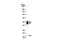
~90 kDa Full Length PDGF D dimer, ~52 kDa PDGF D hemi-dimer,~32 kDa growth factor dimer in the non-reducing conditions; 42.85 kDa calculated. Uncharacterized bands may be observed in some lysate(s).
Physicochemical Information
Dimensions
Materials Information
Toxicological Information
Safety Information according to GHS
Safety Information
Product Usage Statements
Quality Assurance
Evaluated by Western Blotting condition media collected from LNCaP cells transfected with PDGF-D expression vector.
Western Blotting Analysis: 2 µg/mL of this antibody detected PDGF-D in PBS exchanged condition media collected from LNCaP cells transfected with PDGF-D expression vector.
Usage Statement
Unless otherwise stated in our catalog or other company documentation accompanying the product(s), our products are intended for research use only and are not to be used for any other purpose, which includes but is not limited to, unauthorized commercial uses, in vitro diagnostic uses, ex vivo or in vivo therapeutic uses or any type of consumption or application to humans or animals.