Wenn Sie das Fenster schließen, wird Ihre Konfiguration nicht gespeichert, es sei denn, Sie haben Ihren Artikel in die Bestellung aufgenommen oder zu Ihren Favoriten hinzugefügt.
Klicken Sie auf OK, um das MILLIPLEX® MAP-Tool zu schließen oder auf Abbrechen, um zu Ihrer Auswahl zurückzukehren.
Wählen Sie konfigurierbare Panels & Premixed-Kits - ODER - Kits für die zelluläre Signaltransduktion & MAPmates™
Konfigurieren Sie Ihre MILLIPLEX® MAP-Kits und lassen sich den Preis anzeigen.
Konfigurierbare Panels & Premixed-Kits
Unser breites Angebot enthält Multiplex-Panels, für die Sie die Analyten auswählen können, die am besten für Ihre Anwendung geeignet sind. Unter einem separaten Register können Sie das Premixed-Cytokin-Format oder ein Singleplex-Kit wählen.
Kits für die zelluläre Signaltransduktion & MAPmates™
Wählen Sie gebrauchsfertige Kits zur Erforschung gesamter Signalwege oder Prozesse. Oder konfigurieren Sie Ihre eigenen Kits mit Singleplex MAPmates™.
Die folgenden MAPmates™ sollten nicht zusammen analysiert werden: -MAPmates™, die einen unterschiedlichen Assaypuffer erfordern. -Phosphospezifische und MAPmate™ Gesamtkombinationen wie Gesamt-GSK3β und Gesamt-GSK3β (Ser 9). -PanTyr und locusspezifische MAPmates™, z.B. Phospho-EGF-Rezeptor und Phospho-STAT1 (Tyr701). -Mehr als 1 Phospho-MAPmate™ für ein einziges Target (Akt, STAT3). -GAPDH und β-Tubulin können nicht mit Kits oder MAPmates™, die panTyr enthalten, analysiert werden.
.
Bestellnummer
Bestellinformationen
St./Pkg.
Liste
Dieser Artikel wurde zu Ihren Favoriten hinzugefügt.
Wählen Sie bitte Spezies, Panelart, Kit oder Probenart
Um Ihr MILLIPLEX® MAP-Kit zu konfigurieren, wählen Sie zunächst eine Spezies, eine Panelart und/oder ein Kit.
Custom Premix Selecting "Custom Premix" option means that all of the beads you have chosen will be premixed in manufacturing before the kit is sent to you.
Catalogue Number
Ordering Description
Qty/Pack
List
Dieser Artikel wurde zu Ihren Favoriten hinzugefügt.
Spezies
Panelart
Gewähltes Kit
Menge
Bestellnummer
Bestellinformationen
St./Pkg.
Listenpreis
96-Well Plate
Menge
Bestellnummer
Bestellinformationen
St./Pkg.
Listenpreis
Weitere Reagenzien hinzufügen (MAPmates erfordern die Verwendung eines Puffer- und Detektionskits)
Menge
Bestellnummer
Bestellinformationen
St./Pkg.
Listenpreis
48-602MAG
Buffer Detection Kit for Magnetic Beads
1 Kit
Platzsparende Option Kunden, die mehrere Kits kaufen, können ihre Multiplex-Assaykomponenten in Kunststoffbeuteln anstelle von Packungen erhalten, um eine kompaktere Lagerung zu ermöglichen.
Dieser Artikel wurde zu Ihren Favoriten hinzugefügt.
Das Produkt wurde in Ihre Bestellung aufgenommen
Sie können nun ein weiteres Kit konfigurieren, ein Premixed-Kit wählen, zur Kasse gehen oder das Bestell-Tool schließen.
Detect Syntaxin 1 using this Anti-Syntaxin 1 Antibody validated for use in WB, IH.
More>>Detect Syntaxin 1 using this Anti-Syntaxin 1 Antibody validated for use in WB, IH. Less<<
Anti-Syntaxin 1 Antibody: SDB (Sicherheitsdatenblätter), Analysenzertifikate und Qualitätszertifikate, Dossiers, Broschüren und andere verfügbare Dokumente.
CONTROL ANTIGEN: Included free of charge with the antibody is 50 μg of control antigen. The stock solution of the antigen can be made up using 100 μL of PBS. For positive control, in Western blot using 50 ng of protein per Minigel lane. For negative control, preincubate 5-10 μg of fusion protein with 1 μg of antibody for one hour at room temperature. Optimal concentrations must be determined by the end user.
Presentation
Affinity purified immunoglobulin. Lyophilized from phosphate buffered saline, pH 7.4, containing 1% BSA, and 0.025% sodium azide as a preservative. Reconstitute with 200 μL of sterile deionized water. Centrifuge antibody preparation before use (10,000 xg for 5 min).
Detect Syntaxin 1 using this Anti-Syntaxin 1 Antibody validated for use in WB, IH.
Key Applications
Western Blotting
Immunohistochemistry
Application Notes
Western blot: 1:1000 using ECL on rat brain membranes.
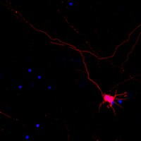
Immunohistochemistry on rat brain sections: AB5820 is directed against an intracellular epitope. Thus, a procedure including permeabilization of cells with 0.2% Triton X 100 is recommended. It is recommended that you start with a working concentration of 30 μg/mL (1:33).
Dilutions should be made using a carrier protein such as BSA (1-3%)
Optimal working dilutions must be determined by the end user.
Biological Information
Immunogen
GST fusion protein corresponding to a cytoplasmic part of rat Syntaxin 1A (Accession number P32851) (Bennet et al. 1992; Inoue & Akagawa 1992).
Concentration
Please refer to the Certificate of Analysis for the lot-specific concentration.
Host
Rabbit
Specificity
Recognizes Syntaxin 1A and Syntaxin 1B. The epitope does not share homology with any other known proteins.
Synaptic vesicles store neurotransmitters that are released during calcium-regulated exocytosis. The specificity of neurotransmitter release requires the localization of both synaptic vesicles and calcium channels to the presynaptic active zone. Syntaxins function in this vesicle fusion process. Syntaxins also serve as a substrate for botulinum neurotoxin type C, a metalloprotease that blocks exocytosis and has high affinity for a molecular complex that includes the alpha-latrotoxin receptor (MIM 600565) which produces explosive exocytosis (Zhang et al., 1995 [PubMed 7622072]).[supplied by OMIM]
FUNCTION: SwissProt: Q16623 # Potentially involved in docking of synaptic vesicles at presynaptic active zones. May play a critical role in neurotransmitter exocytosis. SIZE: 288 amino acids; 33023 Da SUBUNIT: Part of the SNARE core complex containing SNAP25, VAMP2 and STX1A. This complex binds to CPLX1. Binds SYTL4 and STXBP6. Found in a ternary complex with STX1A and SNAP25 (By similarity). Found in a complex with VAMP8 and SNAP23. Interacts with VAPA and SYBU. SUBCELLULAR LOCATION: Cell membrane; Single-pass type IV membrane protein. & Isoform 2: Secreted (Probable). TISSUE SPECIFICITY: Isoform 1 is highly expressed in embryonic spinal chord and ganglia and in adult cerebellum and cerebral cortex. Isoform 2 is expressed in heart, liver, fat, skeletal muscle, kidney and brain. DISEASE: SwissProt: Q16623 # Haploinsufficiency of STX1A may be the cause of certain cardiovascular and musculo-skeletal abnormalities observed in Williams-Beuren syndrome (WBS), a rare developmental disorder. It is a contiguous gene deletion syndrome involving genes from chromosome band 7q11.23. SIMILARITY: SwissProt: Q16623 ## Belongs to the syntaxin family. & Contains 1 t-SNARE coiled-coil homology domain.
Physicochemical Information
Dimensions
Materials Information
Toxicological Information
Safety Information according to GHS
Safety Information
Product Usage Statements
Usage Statement
Unless otherwise stated in our catalog or other company documentation accompanying the product(s), our products are intended for research use only and are not to be used for any other purpose, which includes but is not limited to, unauthorized commercial uses, in vitro diagnostic uses, ex vivo or in vivo therapeutic uses or any type of consumption or application to humans or animals.
Storage and Shipping Information
Storage Conditions
Maintain lyophilized material at -20°C for up to 12 months after date of receipt. After reconstitution maintain at -20°C in undiluted aliquots for up to 6 months. Avoid repeated freeze/thaw cycles.
Syntaxin: a synaptic protein implicated in docking of synaptic vesicles at presynaptic active zones. Bennett, M K, et al. Science, 257: 255-9 (1992)
1992
Neuron-specific antigen HPC-1 from bovine brain reveals strong homology to epimorphin, an essential factor involved in epithelial morphogenesis: identification of a novel protein family. Inoue, A and Akagawa, K Biochem. Biophys. Res. Commun., 187: 1144-50 (1992)
1992
We have already cloned the cDNA for the HPC-1 antigen, a neuron-specific protein antigen from the rat brain. Here we report the molecular cloning of the bovine HPC-1 antigen homologue, and much strong sequence conservation between rat and bovine. By searching the recent protein data base, it was found that the HPC-1 antigen revealed unusual similarity to epimorphin which was mesenchymal factor related to the morphogenesis of primitive epidermal tissues in embryonic stages. We also found that the HPC-1 antigen was identical to p35A (syntaxin) which bound both to a synaptic vesicle protein and to N-type calcium channel. Although the relationship of the physiological functions, structures and topologies along cellular membrane between the HPC-1 antigen and epimorphin have not been consistent yet, these two proteins belong to a novel protein family.
The nervous system coordinates the voluntary and involuntary actions of the individual and transmits signals between different parts of the body. Weitere Informationen >>