Wenn Sie das Fenster schließen, wird Ihre Konfiguration nicht gespeichert, es sei denn, Sie haben Ihren Artikel in die Bestellung aufgenommen oder zu Ihren Favoriten hinzugefügt.
Klicken Sie auf OK, um das MILLIPLEX® MAP-Tool zu schließen oder auf Abbrechen, um zu Ihrer Auswahl zurückzukehren.
Wählen Sie konfigurierbare Panels & Premixed-Kits - ODER - Kits für die zelluläre Signaltransduktion & MAPmates™
Konfigurieren Sie Ihre MILLIPLEX® MAP-Kits und lassen sich den Preis anzeigen.
Konfigurierbare Panels & Premixed-Kits
Unser breites Angebot enthält Multiplex-Panels, für die Sie die Analyten auswählen können, die am besten für Ihre Anwendung geeignet sind. Unter einem separaten Register können Sie das Premixed-Cytokin-Format oder ein Singleplex-Kit wählen.
Kits für die zelluläre Signaltransduktion & MAPmates™
Wählen Sie gebrauchsfertige Kits zur Erforschung gesamter Signalwege oder Prozesse. Oder konfigurieren Sie Ihre eigenen Kits mit Singleplex MAPmates™.
Die folgenden MAPmates™ sollten nicht zusammen analysiert werden: -MAPmates™, die einen unterschiedlichen Assaypuffer erfordern. -Phosphospezifische und MAPmate™ Gesamtkombinationen wie Gesamt-GSK3β und Gesamt-GSK3β (Ser 9). -PanTyr und locusspezifische MAPmates™, z.B. Phospho-EGF-Rezeptor und Phospho-STAT1 (Tyr701). -Mehr als 1 Phospho-MAPmate™ für ein einziges Target (Akt, STAT3). -GAPDH und β-Tubulin können nicht mit Kits oder MAPmates™, die panTyr enthalten, analysiert werden.
.
Bestellnummer
Bestellinformationen
St./Pkg.
Liste
Dieser Artikel wurde zu Ihren Favoriten hinzugefügt.
Wählen Sie bitte Spezies, Panelart, Kit oder Probenart
Um Ihr MILLIPLEX® MAP-Kit zu konfigurieren, wählen Sie zunächst eine Spezies, eine Panelart und/oder ein Kit.
Custom Premix Selecting "Custom Premix" option means that all of the beads you have chosen will be premixed in manufacturing before the kit is sent to you.
Catalogue Number
Ordering Description
Qty/Pack
List
Dieser Artikel wurde zu Ihren Favoriten hinzugefügt.
Spezies
Panelart
Gewähltes Kit
Menge
Bestellnummer
Bestellinformationen
St./Pkg.
Listenpreis
96-Well Plate
Menge
Bestellnummer
Bestellinformationen
St./Pkg.
Listenpreis
Weitere Reagenzien hinzufügen (MAPmates erfordern die Verwendung eines Puffer- und Detektionskits)
Menge
Bestellnummer
Bestellinformationen
St./Pkg.
Listenpreis
48-602MAG
Buffer Detection Kit for Magnetic Beads
1 Kit
Platzsparende Option Kunden, die mehrere Kits kaufen, können ihre Multiplex-Assaykomponenten in Kunststoffbeuteln anstelle von Packungen erhalten, um eine kompaktere Lagerung zu ermöglichen.
Dieser Artikel wurde zu Ihren Favoriten hinzugefügt.
Das Produkt wurde in Ihre Bestellung aufgenommen
Sie können nun ein weiteres Kit konfigurieren, ein Premixed-Kit wählen, zur Kasse gehen oder das Bestell-Tool schließen.
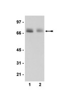
This Anti-phospho-Paxillin (Tyr118) Antibody is validated for use in WB for the detection of phospho-Paxillin (Tyr118).
More>>This Anti-phospho-Paxillin (Tyr118) Antibody is validated for use in WB for the detection of phospho-Paxillin (Tyr118). Less<<
Anti-phospho-Paxillin (Tyr118) Antibody: SDB (Sicherheitsdatenblätter), Analysenzertifikate und Qualitätszertifikate, Dossiers, Broschüren und andere verfügbare Dokumente.
This Anti-phospho-Paxillin (Tyr118) Antibody is validated for use in WB for the detection of phospho-Paxillin (Tyr118).
Key Applications
Western Blotting
Biological Information
Immunogen
KLH-conjugated, synthetic peptide corresponding to amino acids 116-127 (HV[pY]SFPNKQKSA-C) of human Paxillin where [pY] is phosphotyrosine 118. A C-terminal cysteine added for conjugation purposes.
Host
Rabbit
Specificity
phospho-Paxillin (Tyr118).
Isotype
IgG
Species Reactivity
Mouse
Species Reactivity Note
Predicted to cross-react with human, rat, monkey, chicken and canine based on sequence homology.
FUNCTION: SwissProt: P49023 # Cytoskeletal protein involved in actin-membrane attachment at sites of cell adhesion to the extracellular matrix (focal adhesion). SIZE: 591 amino acids; 64533 Da SUBUNIT: Binds in vitro to vinculin as well as to the SH3 domain of c-SRC and, when tyrosine phosphorylated, to the SH2 domain of V-CRK. Isoform beta binds to focal adhesion kinase but weakly to vinculin. Isoform gamma binds to vinculin but only weakly to focal adhesion kinase. Interacts with GIT1, NUDT16L1/SDOS, PARVA and TGFB1I1. Component of cytoplasmic complexes, which also contain GIT1, ARHGEF6 and PAK1 (By similarity). Binds DDEF2. Interacts with unphosphorylated ITGA4. Interacts with RNF5. PTM: Phosphorylated on tyrosine residues during integrin-mediated cell adhesion, embryonic development, fibroblast transformation and following stimulation of cells by mitogens. SIMILARITY: SwissProt: P49023 ## Belongs to the paxillin family. & Contains 4 LIM zinc-binding domains.
Molecular Weight
Mr 68kDa
Physicochemical Information
Dimensions
Materials Information
Toxicological Information
Safety Information according to GHS
Safety Information
Product Usage Statements
Quality Assurance
Routinely evaluated by immunoblot.
Usage Statement
Unless otherwise stated in our catalog or other company documentation accompanying the product(s), our products are intended for research use only and are not to be used for any other purpose, which includes but is not limited to, unauthorized commercial uses, in vitro diagnostic uses, ex vivo or in vivo therapeutic uses or any type of consumption or application to humans or animals.
The NLRP3 Inflammasome Is a Pathogen Sensor for Invasive Entamoeba histolytica via Activation of α5β1 Integrin at the Macrophage-Amebae Intercellular Junction. Mortimer, L; Moreau, F; Cornick, S; Chadee, K PLoS pathogens
11
e1004887
2015
Entamoeba histolytica (Eh) is an extracellular protozoan parasite of humans that invades the colon to cause life-threatening intestinal and extra-intestinal amebiasis. Colonized Eh is asymptomatic, however, when trophozoites adhere to host cells there is a considerable inflammatory response that is critical in the pathogenesis of amebiasis. The host and/or parasite factors that trigger the inflammatory response to invading Eh are not well understood. We recently identified that Eh adherence to macrophages induces inflammasome activation and in the present study we sought to determine the molecular events upon contact that coordinates this response. Here we report that Eh contact-dependent activation of α5β1 integrin is critical for activation of the NLRP3 inflammasome. Eh-macrophage contact triggered recruitment of α5β1 integrin and NLRP3 into the intercellular junction, where α5β1 integrin underwent activation by an integrin-binding cysteine protease on the parasite surface, termed EhCP5. As a result of its activation, α5β1 integrin induced ATP release into the extracellular space through opening of pannexin-1 channels that signalled through P2X7 receptors to deliver a critical co-stimulatory signal that activated the NLRP3 inflammasome. Both the cysteine protease activity and integrin-binding domain of EhCP5 were required to trigger α5β1 integrin that led to ATP release and NLRP3 inflammasome activation. These findings reveal engagement of α5β1 integrin across the parasite-host junction is a key regulatory step that initiates robust inflammatory responses to Eh. We propose that α5β1 integrin distinguishes Eh direct contact and functions with NLRP3 as pathogenicity sensor for invasive Eh infection.
Molecular scaffold or adaptor proteins facilitate precise spatiotemporal regulation and integration of multiple signaling pathways to effect the optimal cellular response to changes in the immediate environment. Paxillin is a multidomain adaptor that recruits both structural and signaling molecules to focal adhesions, sites of integrin engagement with the extracellular matrix, where it performs a critical role in transducing adhesion and growth factor signals to elicit changes in cell migration and gene expression.
Src family nonreceptor protein tyrosine kinases transduce signals that control normal cellular processes such as cell proliferation, adhesion and motility. Normally, cellular Src is held in an inactive state, but in several cancer types, abnormal events lead to elevated kinase activity of the protein and cause pleiotropic cellular responses inducing transformation and metastasis. A prerequisite of the ability of a cancer cell to undergo metastasis into distant tissues is to penetrate surrounding extracellular matrices. These processes are facilitated by the integrin family of cell adhesion molecules. As is the case with Src, altered integrin activity or substrate affinity can contribute to the neoplastic phenotype. Therefore, understanding the interplay between Src and integrin function has been of intense interest over the past few years. This review focuses on the role of Src and integrin signaling in normal cells and how this is deregulated in human cancer. We will identify the key players in the integrin-mediated signaling pathways involved in cell motility and apoptosis, such as FAK, paxillin and p130(CAS), and discuss how Src signaling affects the formation of focal adhesions and the extracellular matrix.
FAK and paxillin are important components in integrin-regulated signaling. New evidence suggests that these two proteins function in crosstalk between cell-matrix and cell-cell adhesions. Further, new insight suggests that under some conditions these proteins inhibit cell migration, in contrast to their established roles in several cell systems as positive regulators of cell adhesion and migration.
Tyrosine phosphorylation of paxillin, FAK, and p130CAS: effects on cell spreading and migration. Panetti, Tracee S Front. Biosci., 7: d143-50 (2002)
2002
Integrins are transmembrane receptors that mediate cell attachment to the substrate. At the cytoplasmic surface of the integrin, cytoskeletal proteins cluster into focal adhesions. The focal adhesions contain multiple proteins that provide a structural and signaling complex inside the cell. This review focuses on three of the cytoskeletal components of the focal adhesion, paxillin, FAK, and p130CAS, that are phosphorylated and play a regulatory role in cell spreading and cell migration. A brief discussion is included of tyrosine phosphorylation of the integrin in relation to localization and phosphorylation of these cytoskeletal proteins. The phosphorylation of integrins and cytoskeletal proteins regulates localization and downstream signaling with profound effects on cell movement.