Development of a highly sensitive immunohistochemical method to detect neurochemical molecules in formalin-fixed and paraffin-embedded tissues from autopsied human brains.
Goto, S; Morigaki, R; Okita, S; Nagahiro, S; Kaji, R
Frontiers in neuroanatomy
9
22
2015
Show Abstract
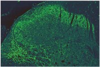
Immunohistochemistry (IHC) is a valuable method for identifying discrete neurochemical molecules by the interaction of target antigens with validated antibodies tagged with a visible label (e.g., peroxidase). We have developed an immunostaining method that is highly sensitive in detection of neurochemical antigens. Our IHC method, which we call the PBTA method, involves a hybrid protocol that implements aspects of both the polymer and avidin-biotin-complex (ABC) methods in combination with biotin-tyramide amplification. When using [Met]-enkephalin as a target antigen, the sensitivity of the PBTA method for IHC was more than 100-fold higher compared with the polymer and ABC methods. In addition, its sensitivity for enzyme-linked immunosorbent assay was about 1,000-fold higher compared with the ABC method. We examined the utility of our IHC method for both chromogenic and fluorescence detection systems used to visualize neurochemical peptides and proteins in formalin-fixed, paraffin-embedded tissues from autopsied human brains. The results convincingly demonstrate that under optimal conditions, our IHC method is highly sensitive without increasing non-specific background activities. Our IHC method could be a powerful tool for detection and visualization of neurochemical antigens that are present even in trace amounts in autopsied human brains. | 25784860
 |
Expression of mu opioid receptor in dorsal diencephalic conduction system: new insights for the medial habenula.
Gardon, O; Faget, L; Chu Sin Chung, P; Matifas, A; Massotte, D; Kieffer, BL
Neuroscience
277
595-609
2014
Show Abstract
The habenular complex, encompassing medial (MHb) and lateral (LHb) divisions, is a highly conserved epithalamic structure involved in the dorsal diencephalic conduction system (DDC). These brain nuclei regulate information flow between the limbic forebrain and the mid- and hindbrain, integrating cognitive with emotional and sensory processes. The MHb is also one of the strongest expression sites for mu opioid receptors (MORs), which mediate analgesic and rewarding properties of opiates. At present however, anatomical distribution and function of these receptors have been poorly studied in MHb pathways. Here we took advantage of a newly generated MOR-mcherry knock-in mouse line to characterize MOR expression sites in the DDC. MOR-mcherry fluorescent signal is weak in the LHb, but strong expression is visible in the MHb, fasciculus retroflexus (fr) and interpeduncular nucleus (IPN), indicating that MOR is mainly present in the MHb-IPN pathway. MOR-mcherry cell bodies are detected both in basolateral and apical parts of MHb, where the receptor co-localizes with cholinergic and substance P (SP) neurons, respectively, representing two main MHb neuronal populations. MOR-mcherry is expressed in most MHb-SP neurons, and is present in only a subpopulation of MHb-cholinergic neurons. Intense diffuse fluorescence detected in lateral and rostral parts of the IPN further suggests that MOR-mcherry is transported to terminals of these SP and cholinergic neurons. Finally, MOR-mcherry is present in septal regions projecting to the MHb, and in neurons of the central and intermediate IPN. Together, this study describes MOR expression in several compartments of the MHb-IPN circuitry. The remarkably high MOR density in the MHb-IPN pathway suggests that these receptors are in a unique position to mediate analgesic, autonomic and reward responses. | 25086313
|
GABAergic inputs from direct and indirect striatal projection neurons onto cholinergic interneurons in the primate putamen.
Gonzales, KK; Pare, JF; Wichmann, T; Smith, Y
The Journal of comparative neurology
521
2502-22
2013
Show Abstract
Striatal cholinergic interneurons (ChIs) are involved in reward-dependent learning and the regulation of attention. The activity of these neurons is modulated by intrinsic and extrinsic γ-aminobutyric acid (GABA)ergic and glutamatergic afferents, but the source and relative prevalence of these diverse regulatory inputs remain to be characterized. To address this issue, we performed a quantitative ultrastructural analysis of the GABAergic and glutamatergic innervation of ChIs in the postcommissural putamen of rhesus monkeys. Postembedding immunogold localization of GABA combined with peroxidase immunostaining for choline acetyltransferase showed that 60% of all synaptic inputs to ChIs originate from GABAergic terminals, whereas 21% are from putatively glutamatergic terminals that establish asymmetric synapses, and 19% from other (non-GABAergic) sources of symmetric synapses. Double pre-embedding immunoelectron microscopy using substance P and Met-/Leu-enkephalin antibodies to label GABAergic terminals from collaterals of "direct" and "indirect" striatal projection neurons, respectively, revealed that 47% of the indirect pathway terminals and 36% of the direct pathway terminals target ChIs. Together, substance P- and enkephalin-positive terminals represent 24% of all synapses onto ChIs in the monkey putamen. These findings show that ChIs receive prominent GABAergic inputs from multiple origins, including a significant contingent from axon collaterals of direct and indirect pathway projection neurons. | 23296794
|
An anxiolytic role for CRF receptor type 1 in the globus pallidus.
Sztainberg, Y; Kuperman, Y; Justice, N; Chen, A
The Journal of neuroscience : the official journal of the Society for Neuroscience
31
17416-24
2011
Show Abstract
Corticotropin-releasing factor receptor type 1 (CRFR1) plays a major role in the regulation of neuroendocrine and behavioral responses to stress and is considered a key mediator of anxiety behavior. The globus pallidus external (GPe), a main relay center within the basal ganglia that is primarily associated with motor and associative functions, is one of the brain nuclei with the highest levels of CRFR1 expression in the rodent brain. However, the role of CRFR1 in the GPe is yet unknown. In the present study, we used a lentiviral-based system of RNA interference to show that knockdown of CRFR1 mRNA expression in the GPe of adult mice induces a significant increase in anxiety-like behavior, as revealed by the light-dark transfer, open-field, and elevated plus-maze tests. This effect was further confirmed by pharmacological administration of the selective CRFR1 antagonist NBI 30775 (1.75 μg/side) directly into the GPe. In the marble-burying test, blockade of CRFR1 in the GPe increased the percentage of marbles buried and the duration of burying behavior. Additionally, we present evidence suggesting that the enkephalin system is involved in the effect of GPe-CRFR1 on anxiety-like behavior. In contrast to the well established anxiogenic role of CRFR1 in the extended amygdala, our data reveal a novel anxiolytic role for CRFR1 in the GPe. | 22131403
|
Loss of Goosecoid-like and DiGeorge syndrome critical region 14 in interpeduncular nucleus results in altered regulation of rapid eye movement sleep.
Funato, H; Sato, M; Sinton, CM; Gautron, L; Williams, SC; Skach, A; Elmquist, JK; Skoultchi, AI; Yanagisawa, M
Proceedings of the National Academy of Sciences of the United States of America
107
18155-60
2010
Show Abstract
Sleep and wakefulness are regulated primarily by inhibitory interactions between the hypothalamus and brainstem. The expression of the states of rapid eye movement (REM) sleep and non-REM (NREM) sleep also are correlated with the activity of groups of REM-off and REM-on neurons in the dorsal brainstem. However, the contribution of ventral brainstem nuclei to sleep regulation has been little characterized to date. Here we examined sleep and wakefulness in mice deficient in a homeobox transcription factor, Goosecoid-like (Gscl), which is one of the genes deleted in DiGeorge syndrome or 22q11 deletion syndrome. The expression of Gscl is restricted to the interpeduncular nucleus (IP) in the ventral region of the midbrain-hindbrain transition. The IP has reciprocal connections with several cell groups implicated in sleep/wakefulness regulation. Although Gscl(-/-) mice have apparently normal anatomy and connections of the IP, they exhibited a reduced total time spent in REM sleep and fewer REM sleep episodes. In addition, Gscl(-/-) mice showed reduced theta power during REM sleep and increased arousability during REM sleep. Gscl(-/-) mice also lacked the expression of DiGeorge syndrome critical region 14 (Dgcr14) in the IP. These results indicate that the absence of Gscl and Dgcr14 in the IP results in altered regulation of REM sleep. Full Text Article | 20921407
|
Differential regulation of morphine antinociceptive effects by endogenous enkephalinergic system in the forebrain of mice.
Chen, TC; Cheng, YY; Sun, WZ; Shyu, BC
Molecular pain
4
41
2008
Show Abstract
Mice lacking the preproenkephalin (ppENK) gene are hyperalgesic and show more anxiety and aggression than wild-type (WT) mice. The marked behavioral changes in ppENK knock-out (KO) mice appeared to occur in supraspinal response to painful stimuli. However the functional role of enkephalins in the supraspinal nociceptive processing and their underlying mechanism is not clear. The aim of present study was to compare supraspinal nociceptive and morphine antinociceptive responses between WT and ppENK KO mice.The genotypes of bred KO mice were confirmed by PCR. Met-enkephalin immunoreactive neurons were labeled in the caudate-putamen, intermediated part of lateral septum, lateral globus pallidus, intermediated part of lateral septum, hypothalamus, and amygdala of WT mice. Met-enkephalin immunoreactive neurons were not found in the same brain areas in KO mice. Tail withdrawal and von Frey test results did not differ between WT and KO mice. KO mice had shorter latency to start paw licking than WT mice in the hot plate test. The maximal percent effect of morphine treatments (5 mg/kg and 10 mg/kg, i.p.) differed between WT and KO mice in hot plate test. The current source density (CSD) profiles evoked by peripheral noxious stimuli in the primary somatosenstory cortex (S1) and anterior cingulate cortex (ACC) were similar in WT and KO mice. After morphine injection, the amplitude of the laser-evoked sink currents was decreased in S1 while the amplitude of electrical-evoked sink currents was increased in the ACC. These differential morphine effects in S1 and ACC were enhanced in KO mice. Facilitation of synaptic currents in the ACC is mediated by GABA inhibitory interneurons in the local circuitry. Percent increases in opioid receptor binding in S1 and ACC were 5.1% and 5.8%, respectively.The present results indicate that the endogenous enkephalin system is not involved in acute nociceptive transmission in the spinal cord, S1, and ACC. However, morphine preferentially suppressed supraspinal related nociceptive behavior in KO mice. This effect was reflected in the potentiated differential effects of morphine in the S1 and ACC in KO mice. This potentiation may be due to an up-regulation of opioid receptors. Thus these findings strongly suggest an antagonistic interaction between the endogenous enkephalinergic system and exogenous opioid analgesic actions in the supraspinal brain structures. | 18826595
|
Photic induction of c-Fos in enkephalin neurons of the rat intergeniculate leaflet innervated by retinal PACAP fibres.
Fie Juhl,Jens Hannibal,Jan Fahrenkrug
Cell and tissue research
329
2007
Show Abstract
The brain's biological clock, located in the suprachiasmatic nucleus (SCN), is synchronised with the cyclic environment by photic and non-photic cues. Photic information to the SCN is mediated by pituitary adenylate-cyclase-activating polypeptide (PACAP)-containing retinal ganglion cells (RGCs), whereas non-photic input originates primarily from neuropeptide Y (NPY) cells in the ipsilateral thalamic intergeniculate leaflet (IGL). RGCs also seem to project to the IGL, indicating a role for this structure in the integration of photic and non-photic inputs related to the resetting of the biological clock. In the present study, we have used anterograde tracing from both eyes, bilateral eye enucleation, double-immunofluorescence histochemistry, high-resolution confocal laser scanning microscopy and three-dimensional computer analysis to show that (1) PACAP-containing RGCs project to the IGL and are the only source for the PACAP-immunoreactive fibres in the IGL; (2) a few NPY-containing neurons in the IGL are innervated by PACAP-containing retinal nerve fibres and the contacts are both axodendritic and axosomatic; (3) most enkephalin-immunoreactive neurons in the IGL are innervated by PACAP-containing retinal afferents and the contacts are mainly axodendritic; (4) light stimulation at various time points activates (as evidenced by c-Fos induction) enkephalin-positive neurons but not NPY-immunoreactive neurons. The findings suggest that PACAP-immunoreactive retinal afferents in the IGL primarily innervate enkephalin-immunoactive neurons and that the enkephalin-containing neurons, which project locally and to the contralateral IGL, are activated by light independent of diurnal time. | 17503087
|
Expression of protein kinase C-substrate mRNAs in the basal ganglia of adult and infant macaque monkeys.
Noriyuki Higo, Takao Oishi, Akiko Yamashita, Yumi Murata, Keiji Matsuda, Motoharu Hayashi
The Journal of comparative neurology
499
662-76
2006
Show Abstract
We performed in situ hybridization histochemistry on the monkey basal ganglia to investigate the mRNA localization of three protein kinase C substrates (GAP-43, MARCKS, and neurogranin), of which expression plays a role in structural changes in neurites and synapses. Weak hybridization signals for GAP-43 mRNA and intense signals for both MARCKS and neurogranin mRNAs were observed in the adult neostriatum. All three of the mRNAs were expressed in both substance P-positive direct pathway neurons and enkephalin-positive indirect pathway neurons. In the nucleus accumbens, the hybridization signals for the three mRNAs were weaker than those in the neostriatum. Double-label in situ hybridization histochemistry in the neostriatum revealed that GAP-43 and neurogranin mRNAs were expressed in a subset of MARCKS-positive neurons. While intense hybridization signals for MARCKS mRNA were observed in all of the other basal ganglia regions such as the globus pallidus, substantia innominata, subthalamic nucleus, and substantia nigra, intense signals for GAP-43 mRNA were restricted to the substantia innominata and substantia nigra pars compacta. No signal for neurogranin mRNA was observed in the basal ganglia regions outside the neostriatum and the nucleus accumbens. These results indicate that the protein kinase C substrates are abundant in some specific connections in cortico-basal ganglia circuits. Developmental analysis showed that the expression level in the putamen and nucleus accumbens, but not in the caudate nucleus, was higher in the infant than in the adult, suggesting that synaptic maturation in the caudate nucleus occurs earlier than that in the putamen and nucleus accumbens. | 17029258
|
Huntingtin distribution among striatal output neurons of normal rat brain
Fusco, F.R. et al.
Neuroscience Letters , 339:53-56 (2003)
2003
| 12618299
|
Neuropeptides.
Moore, M R and Black, P M
Neurosurgical review, 14: 97-110 (1991)
1991
Show Abstract
This review summarizes the revolutionary impact of brain peptides on our understanding of the nervous system and then discusses the localization, distribution, synthesis, receptor sites, and possible function of 32 brain peptides. The peptides are discussed in three subgroups: I) the opioid peptides, which include beta-endorphin, the enkephalins, and dynorphin; II) the pituitary releasing hormones, most of which are wide-spread in the brain and include corticotropin-releasing hormone, luteinizing hormone-releasing hormone, somatostatin, and thyrotropin-releasing hormone; and III) a selection of 12 other peptides potentially important for neurological function, including vasopressin, oxytocin, substance P, cholecystokinin, bombesin, neurotensin, renin, angiotensin, vasoactive intestinal polypeptide, neuropeptide Y, calcitonin gene-related peptide, and calcitonin. Within each individual peptide section, the possible physiological roles in anterior pituitary hormone release, blood-flow regulation, feeding behavior, temperature regulation, nociception, memory and learning, and movement are reviewed. Further, where noted, the peptide findings in Huntington's, Alzheimer's, Parkinson's and psychiatric diseases are emphasized. | 1870724
|