Chd1 is essential for the high transcriptional output and rapid growth of the mouse epiblast.
Guzman-Ayala, M; Sachs, M; Koh, FM; Onodera, C; Bulut-Karslioglu, A; Lin, CJ; Wong, P; Nitta, R; Song, JS; Ramalho-Santos, M
Development (Cambridge, England)
142
118-27
2015
Show Abstract
The pluripotent mammalian epiblast undergoes unusually fast cell proliferation. This rapid growth is expected to generate a high transcriptional demand, but the underlying mechanisms remain unknown. We show here that the chromatin remodeler Chd1 is required for transcriptional output and development of the mouse epiblast. Chd1(-/-) embryos exhibit proliferation defects and increased apoptosis, are smaller than controls by E5.5 and fail to grow, to become patterned or to gastrulate. Removal of p53 allows progression of Chd1(-/-) mutants only to E7.0-8.0, highlighting the crucial requirement for Chd1 during early post-implantation development. Chd1(-/-) embryonic stem cells (ESCs) have a self-renewal defect and a genome-wide reduction in transcriptional output at both known mRNAs and intergenic transcripts. These transcriptional defects were only uncovered when cell number-normalized approaches were used, and correlate with a lower engagement of RNAP II with transcribed genes in Chd1(-/-) ESCs. We further show that Chd1 directly binds to ribosomal DNA, and that both Chd1(-/-) epiblast cells in vivo and ESCs in vitro express significantly lower levels of ribosomal RNA. In agreement with these findings, mutant cells in vivo and in vitro exhibit smaller and more elongated nucleoli. Thus, the RNA output by both Pol I and II is reduced in Chd1(-/-) cells. Our data indicate that Chd1 promotes a globally elevated transcriptional output required to sustain the distinctly rapid growth of the mouse epiblast. | | | 25480920
 |
Alternative meiotic chromatid segregation in the holocentric plant Luzula elegans.
Heckmann, S; Jankowska, M; Schubert, V; Kumke, K; Ma, W; Houben, A
Nature communications
5
4979
2014
Show Abstract
Holocentric chromosomes occur in a number of independent eukaryotic lineages. They form holokinetic kinetochores along the entire poleward chromatid surfaces, and owing to this alternative chromosome structure, species with holocentric chromosomes cannot use the two-step loss of cohesion during meiosis typical for monocentric chromosomes. Here we show that the plant Luzula elegans maintains a holocentric chromosome architecture and behaviour throughout meiosis, and in contrast to monopolar sister centromere orientation, the unfused holokinetic sister centromeres behave as two distinct functional units during meiosis I, resulting in sister chromatid separation. Homologous non-sister chromatids remain terminally linked after metaphase I, by satellite DNA-enriched chromatin threads, until metaphase II. They then separate at anaphase II. Thus, an inverted sequence of meiotic sister chromatid segregation occurs. This alternative meiotic process is most likely one possible adaptation to handle a holocentric chromosome architecture and behaviour during meiosis. | | | 25296379
|
Dopamine signaling leads to loss of Polycomb repression and aberrant gene activation in experimental parkinsonism.
Södersten, E; Feyder, M; Lerdrup, M; Gomes, AL; Kryh, H; Spigolon, G; Caboche, J; Fisone, G; Hansen, K
PLoS genetics
10
e1004574
2014
Show Abstract
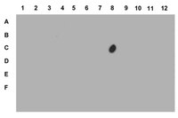
Polycomb group (PcG) proteins bind to and repress genes in embryonic stem cells through lineage commitment to the terminal differentiated state. PcG repressed genes are commonly characterized by the presence of the epigenetic histone mark H3K27me3, catalyzed by the Polycomb repressive complex 2. Here, we present in vivo evidence for a previously unrecognized plasticity of PcG-repressed genes in terminally differentiated brain neurons of parkisonian mice. We show that acute administration of the dopamine precursor, L-DOPA, induces a remarkable increase in H3K27me3S28 phosphorylation. The induction of the H3K27me3S28p histone mark specifically occurs in medium spiny neurons expressing dopamine D1 receptors and is dependent on Msk1 kinase activity and DARPP-32-mediated inhibition of protein phosphatase-1. Chromatin immunoprecipitation (ChIP) experiments showed that increased H3K27me3S28p was accompanied by reduced PcG binding to regulatory regions of genes. An analysis of the genome wide distribution of L-DOPA-induced H3K27me3S28 phosphorylation by ChIP sequencing (ChIP-seq) in combination with expression analysis by RNA-sequencing (RNA-seq) showed that the induction of H3K27me3S28p correlated with increased expression of a subset of PcG repressed genes. We found that induction of H3K27me3S28p persisted during chronic L-DOPA administration to parkisonian mice and correlated with aberrant gene expression. We propose that dopaminergic transmission can activate PcG repressed genes in the adult brain and thereby contribute to long-term maladaptive responses including the motor complications, or dyskinesia, caused by prolonged administration of L-DOPA in Parkinson's disease. | Western Blotting | Mouse | 25254549
|
Genome-wide phosphoacetylation of histone H3 at Drosophila enhancers and promoters.
Kellner, WA; Ramos, E; Van Bortle, K; Takenaka, N; Corces, VG
Genome research
22
1081-8
2012
Show Abstract
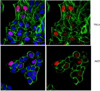
Transcription regulation is mediated by enhancers that bind sequence-specific transcription factors, which in turn interact with the promoters of the genes they control. Here, we show that the JIL-1 kinase is present at both enhancers and promoters of ecdysone-induced Drosophila genes, where it phosphorylates the Ser10 and Ser28 residues of histone H3. JIL-1 is also required for CREB binding protein (CBP)-mediated acetylation of Lys27, a well-characterized mark of active enhancers. The presence of these proteins at enhancers and promoters of ecdysone-induced genes results in the establishment of the H3K9acS10ph and H3K27acS28ph marks at both regulatory sequences. These modifications are necessary for the recruitment of 14-3-3, a scaffolding protein capable of facilitating interactions between two simultaneously bound proteins. Chromatin conformation capture assays indicate that interaction between the enhancer and the promoter is dependent on the presence of JIL-1, 14-3-3, and CBP. Genome-wide analyses extend these conclusions to most Drosophila genes, showing that the presence of JIL-1, H3K9acS10ph, and H3K27acS28ph is a general feature of enhancers and promoters in this organism. | | | 22508764
|
Histone H3 phosphorylation (Ser10, Ser28) and phosphoacetylation (K9S10) are differentially associated with gene expression in liver of rats treated in vivo with acute ethanol.
James, TT; Aroor, AR; Lim, RW; Shukla, SD
J Pharmacol Exp Ther
340
237-47
2012
Show Abstract
The epigenetic histone modification by ethanol is emerging as one of the mechanisms for its deleterious effects in the liver. In this context, we have investigated the role of histone H3 phosphorylation at Ser10 (P-H3-Ser10), and Ser28 (P-H3-Ser28) in liver after acute ethanol treatment in vivo. Ethanol was administered intraperitoneally in male Sprague-Dawley rats. Ethanol dose-response (1-5 g/kg body weight) and time-course (1-4 h) experiments were conducted, and various parameters were monitored. Steatosis and necrosis (serum alanine aminotransferase) of the liver increased in 4 h, suggesting liver injury. There were differences between P-H3-Ser10 and P-H3-Ser28 at 1 h, with the latter being more sensitive to lower ethanol doses. It was noteworthy that phosphorylation of both serines disappeared at the highest dose used (5 g/kg). We also examined phosphoacetylation of histone H3 at K9S10 and observed a dramatic increase. The changes in histone H3 phosphorylation and phosphoacetylation were also accompanied with expression of early response genes (c-fos, c-jun, mitogen-activated protein kinase phosphatase-1). Chromatin immunoprecipitation assays in samples from 1.5 and 4 h of ethanol administration indicated that increased histone H3 phosphorylation at Ser28 was associated with the promoters of c-jun and plasminogen activator inhibitor-1. In conclusion, this study demonstrates for the first time that in vivo exposure of liver to acute ethanol induced phosphorylation and phosphoacetylation of histone H3, and these modifications are differentially involved in the mRNA expression of genes. | | | 22025646
|
Calpain-1 cleaves Rad21 to promote sister chromatid separation.
Panigrahi, AK; Zhang, N; Mao, Q; Pati, D
Molecular and cellular biology
31
4335-47
2011
Show Abstract
Defining the mechanisms of chromosomal cohesion and dissolution of the cohesin complex from chromatids is important for understanding the chromosomal missegregation seen in many tumor cells. Here we report the identification of a novel cohesin-resolving protease and describe its role in chromosomal segregation. Sister chromatids are held together by cohesin, a multiprotein ring-like complex comprised of Rad21, Smc1, Smc3, and SA2 (or SA1). Cohesin is known to be removed from vertebrate chromosomes by two distinct mechanisms, namely, the prophase and anaphase pathways. First, PLK1-mediated phosphorylation of SA2 in prophase leads to release of cohesin from chromosome arms, leaving behind centromeric cohesins that continue to hold the sisters together. Then, at the onset of anaphase, activated separase cleaves the centromeric cohesin Rad21, thereby opening the cohesin ring and allowing the sister chromatids to separate. We report here that the calcium-dependent cysteine endopeptidase calpain-1 is a Rad21 peptidase and normally localizes to the interphase nuclei and chromatin. Calpain-1 cleaves Rad21 at L192, in a calcium-dependent manner. We further show that Rad21 cleavage by calpain-1 promotes separation of chromosome arms, which coincides with a calcium-induced partial loss of cohesin at several chromosomal loci. Engineered cleavage of Rad21 at the calpain-cleavable site without activation of calpain-1 can lead to a loss of sister chromatid cohesion. Collectively, our work reveals a novel function of calpain-1 and describes an additional pathway for sister chromatid separation in humans. | | | 21876002
|
Role of anti-angiogenic factor endostatin in the pathogenesis of experimental ulcerative colitis.
Tolstanova G, Deng X, Khomenko T, Garg P, Paunovic B, Chen L, Sitaraman SV, Shiloach J, Szabo S, Sandor Z.
Life sciences
88
74-81
2011
Show Abstract
Vascular endothelial growth factor (VEGF) and pathologic angiogenesis have been demonstrated to play a pathogenic role in the development and progression of inflammatory bowel disease. Thus, we hypothesized that the potent anti-angiogenic factor endostatin might play a beneficial role in experimental ulcerative colitis (UC). | | | 21047522
|
Chromatin regulated interchange between polycomb repressive complex 2 (PRC2)-Ezh2 and PRC2-Ezh1 complexes controls myogenin activation in skeletal muscle cells.
Stojic, L; Jasencakova, Z; Prezioso, C; Stützer, A; Bodega, B; Pasini, D; Klingberg, R; Mozzetta, C; Margueron, R; Puri, PL; Schwarzer, D; Helin, K; Fischle, W; Orlando, V
Epigenetics & chromatin
4
16
2011
Show Abstract
Polycomb group (PcG) genes code for chromatin multiprotein complexes that are responsible for maintaining gene silencing of transcriptional programs during differentiation and in adult tissues. Despite the large amount of information on PcG function during development and cell identity homeostasis, little is known regarding the dynamics of PcG complexes and their role during terminal differentiation.We show that two distinct polycomb repressive complex (PRC)2 complexes contribute to skeletal muscle cell differentiation: the PRC2-Ezh2 complex, which is bound to the myogenin (MyoG) promoter and muscle creatine kinase (mCK) enhancer in proliferating myoblasts, and the PRC2-Ezh1 complex, which replaces PRC2-Ezh2 on MyoG promoter in post-mitotic myotubes. Interestingly, the opposing dynamics of PRC2-Ezh2 and PRC2-Ezh1 at these muscle regulatory regions is differentially regulated at the chromatin level by Msk1 dependent methyl/phospho switch mechanism involving phosphorylation of serine 28 of the H3 histone (H3S28ph). While Msk1/H3S28ph is critical for the displacement of the PRC2-Ezh2 complex, this pathway does not influence the binding of PRC2-Ezh1 on the chromatin. Importantly, depletion of Ezh1 impairs muscle differentiation and the chromatin recruitment of MyoD to the MyoG promoter in differentiating myotubes. We propose that PRC2-Ezh1 is necessary for controlling the proper timing of MyoG transcriptional activation and thus, in contrast to PRC2-Ezh2, is required for myogenic differentiation.Our data reveal another important layer of epigenetic control orchestrating skeletal muscle cell terminal differentiation, and introduce a novel function of the PRC2-Ezh1 complex in promoter setting. | | | 21892963
|
Coordination of cell proliferation and anterior-posterior axis establishment in the mouse embryo.
Stuckey, DW; Clements, M; Di-Gregorio, A; Senner, CE; Le Tissier, P; Srinivas, S; Rodriguez, TA
Development (Cambridge, England)
138
1521-30
2011
Show Abstract
During development, the growth of the embryo must be coupled to its patterning to ensure correct and timely morphogenesis. In the mouse embryo, migration of the anterior visceral endoderm (AVE) to the prospective anterior establishes the anterior-posterior (A-P) axis. By analysing the distribution of cells in S phase, M phase and G2 from the time just prior to the migration of the AVE until 18 hours after its movement, we show that there is no evidence for differential proliferation along the A-P axis of the mouse embryo. Rather, we have identified that as AVE movements are being initiated, the epiblast proliferates at a much higher rate than the visceral endoderm. We show that these high levels of proliferation in the epiblast are dependent on Nodal signalling and are required for A-P establishment, as blocking cell division in the epiblast inhibits AVE migration. Interestingly, inhibition of migration by blocking proliferation can be rescued by Dkk1. This suggests that the high levels of epiblast proliferation function to move the prospective AVE away from signals that are inhibitory to its migration. The finding that initiation of AVE movements requires a certain level of proliferation in the epiblast provides a mechanism whereby A-P axis development is coordinated with embryonic growth. | | | 21427142
|
Coe genes are expressed in differentiating neurons in the central nervous system of protostomes.
Demilly, A; Simionato, E; Ohayon, D; Kerner, P; Garcès, A; Vervoort, M
PloS one
6
e21213
2011
Show Abstract
Genes of the coe (collier/olfactory/early B-cell factor) family encode Helix-Loop-Helix transcription factors that are widely conserved in metazoans and involved in many developmental processes, neurogenesis in particular. Whereas their functions during vertebrate neural tube formation have been well documented, very little is known about their expression and role during central nervous system (CNS) development in protostomes. Here we characterized the CNS expression of coe genes in the insect Drosophila melanogaster and the polychaete annelid Platynereis dumerilii, which belong to different subgroups of protostomes and show strikingly different modes of development. In the Drosophila ventral nerve cord, we found that the Collier-expressing cells form a subpopulation of interneurons with diverse molecular identities and neurotransmitter phenotypes. We also demonstrate that collier is required for the proper differentiation of some interneurons belonging to the Eve-Lateral cluster. In Platynereis dumerilii, we cloned a single coe gene, Pdu-coe, and found that it is exclusively expressed in post mitotic neural cells. Using an original technique of in silico 3D registration, we show that Pdu-coe is co-expressed with many different neuronal markers and therefore that, like in Drosophila, its expression defines a heterogeneous population of neurons with diverse molecular identities. Our detailed characterization and comparison of coe gene expression in the CNS of two distantly-related protostomes suggest conserved roles of coe genes in neuronal differentiation in this clade. As similar roles have also been observed in vertebrates, this function was probably already established in the last common ancestor of all bilaterians. | | | 21695052
|