Dendritic Cell-Mediated Phagocytosis but Not Immune Activation Is Enhanced by Plasmin.
Borg, RJ; Samson, AL; Au, AE; Scholzen, A; Fuchsberger, M; Kong, YY; Freeman, R; Mifsud, NA; Plebanski, M; Medcalf, RL
PloS one
10
e0131216
2015
Mostrar resumen
Removal of dead cells in the absence of concomitant immune stimulation is essential for tissue homeostasis. We recently identified an injury-induced protein misfolding event that orchestrates the plasmin-dependent proteolytic degradation of necrotic cells. As impaired clearance of dead cells by the innate immune system predisposes to autoimmunity, we determined whether plasmin could influence endocytosis and immune cell stimulation by dendritic cells - a critical cell that links the innate and adaptive immune systems. We find that plasmin generated on the surface of necrotic cells enhances their phagocytic removal by human monocyte-derived dendritic cells. Plasmin also promoted phagocytosis of protease-resistant microparticles by diverse mouse dendritic cell sub-types both in vitro and in vivo. Together with an increased phagocytic capacity, plasmin-treated dendritic cells maintain an immature phenotype, exhibit reduced migration to lymph nodes, increase their expression/release of the immunosuppressive cytokine TGF-β, and lose their capacity to mount an allogeneic response. Collectively, our findings support a novel role for plasmin formed on dead cells and other phagocytic targets in maintaining tissue homeostasis by increasing the phagocytic function of dendritic cells while simultaneously decreasing their immunostimulatory capacity consistent with producing an immunosuppressive state. | | | 26132730
 |
Reliable LC3 and p62 autophagy marker detection in formalin fixed paraffin embedded human tissue by immunohistochemistry.
Schläfli, AM; Berezowska, S; Adams, O; Langer, R; Tschan, MP
European journal of histochemistry : EJH
59
2481
2015
Mostrar resumen
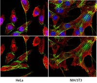
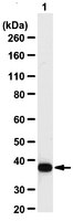
Autophagy assures cellular homeostasis, and gains increasing importance in cancer, where it impacts on carcinogenesis, propagation of the malignant phenotype and development of resistance. To date, its tissue-based analysis by immunohistochemistry remains poorly standardized. Here we show the feasibility of specifically and reliably assessing the autophagy markers LC3B and p62 (SQSTM1) in formalin fixed and paraffin embedded human tissue by immunohistochemistry. Preceding functional experiments consisted of depleting LC3B and p62 in H1299 lung cancer cells with subsequent induction of autophagy. Western blot and immunofluorescence validated antibody specificity, knockdown efficiency and autophagy induction prior to fixation in formalin and embedding in paraffin. LC3B and p62 antibodies were validated on formalin fixed and paraffin embedded cell pellets of treated and control cells and finally applied on a tissue microarray with 80 human malignant and non-neoplastic lung and stomach formalin fixed and paraffin embedded tissue samples. Dot-like staining of various degrees was observed in cell pellets and 18/40 (LC3B) and 22/40 (p62) tumors, respectively. Seventeen tumors were double positive for LC3B and p62. P62 displayed additional significant cytoplasmic and nuclear staining of unknown significance. Interobserver-agreement for grading of staining intensities and patterns was substantial to excellent (kappa values 0.60 - 0.83). In summary, we present a specific and reliable IHC staining of LC3B and p62 on formalin fixed and paraffin embedded human tissue. Our presented protocol is designed to aid reliable investigation of dysregulated autophagy in solid tumors and may be used on large tissue collectives. | | | 26150155
|
Mitofusin 2 Deficiency Affects Energy Metabolism and Mitochondrial Biogenesis in MEF Cells.
Kawalec, M; Boratyńska-Jasińska, A; Beręsewicz, M; Dymkowska, D; Zabłocki, K; Zabłocka, B
PloS one
10
e0134162
2015
Mostrar resumen
Mitofusin 2 (Mfn2), mitochondrial outer membrane protein which is involved in rearrangement of these organelles, was first described in pathology of hypertension and diabetes, and more recently much attention is paid to its functions in Charcot-Marie-Tooth type 2A neuropathy (CMT2A). Here, cellular energy metabolism was investigated in mouse embryonic fibroblasts (MEF) differing in the presence of the Mfn2 gene; control (MEFwt) and with Mfn2 gene depleted MEFMfn2-/-. These two cell lines were compared in terms of various parameters characterizing mitochondrial bioenergetics. Here, we have shown that relative rate of proliferation of MEFMfn2-/- cells versus control fibroblasts depend on serum supplementation of the growth media. Moreover, MEFMfn2-/- cells exhibited significantly increased respiration rate in comparison to MEFwt, regardless of serum supplementation of the medium. This effect was correlated with increased level of mitochondrial markers (TOM20 and NAO) as well as mitochondrial transcription factor A (TFAM) and peroxisome proliferator-activated receptor gamma coactivator 1-alpha (PGC-1α) protein levels and unchanged total ATP content. Interestingly, mitochondrial DNA content in MEFMfn2-/- cells was not reduced. Fundamentally, these results are in contrast to a commonly accepted belief that mitofusin 2 deficiency inevitably results in debilitation of mitochondrial energy metabolism. However, we suggest a balance between negative metabolic consequences of mitofusin 2 deficiency and adaptive processes exemplified by increased level of PGC-1α and TFAM transcription factor which prevent an excessive depletion of mtDNA and severe impairment of cell metabolism. | | | 26230519
|
Impaired spatial memory and enhanced long-term potentiation in mice with forebrain-specific ablation of the Stim genes.
Garcia-Alvarez, G; Shetty, MS; Lu, B; Yap, KA; Oh-Hora, M; Sajikumar, S; Bichler, Z; Fivaz, M
Frontiers in behavioral neuroscience
9
180
2015
Mostrar resumen
Recent findings point to a central role of the endoplasmic reticulum-resident STIM (Stromal Interaction Molecule) proteins in shaping the structure and function of excitatory synapses in the mammalian brain. The impact of the Stim genes on cognitive functions remains, however, poorly understood. To explore the function of the Stim genes in learning and memory, we generated three mouse strains with conditional deletion (cKO) of Stim1 and/or Stim2 in the forebrain. Stim1, Stim2, and double Stim1/Stim2 cKO mice show no obvious brain structural defects or locomotor impairment. Analysis of spatial reference memory in the Morris water maze revealed a mild learning delay in Stim1 cKO mice, while learning and memory in Stim2 cKO mice was indistinguishable from their control littermates. Deletion of both Stim genes in the forebrain resulted, however, in a pronounced impairment in spatial learning and memory reflecting a synergistic effect of the Stim genes on the underlying neural circuits. Notably, long-term potentiation (LTP) at CA3-CA1 hippocampal synapses was markedly enhanced in Stim1/Stim2 cKO mice and was associated with increased phosphorylation of the AMPA receptor subunit GluA1, the transcriptional regulator CREB and the L-type Voltage-dependent Ca(2+) channel Cav1.2 on protein kinase A (PKA) sites. We conclude that STIM1 and STIM2 are key regulators of PKA signaling and synaptic plasticity in neural circuits encoding spatial memory. Our findings also reveal an inverse correlation between LTP and spatial learning/memory and suggest that abnormal enhancement of cAMP/PKA signaling and synaptic efficacy disrupts the formation of new memories. | | | 26236206
|
Protease Omi facilitates neurite outgrowth in mouse neuroblastoma N2a cells by cleaving transcription factor E2F1.
Ma, Q; Hu, QS; Xu, RJ; Zhen, XC; Wang, GH
Acta pharmacologica Sinica
36
966-75
2015
Mostrar resumen
Omi is an ATP-independent serine protease that is necessary for neuronal function and survival. The aim of this study was to investigate the role of protease Omi in regulating differentiation of mouse neuroblastoma cells and to identify the substrate of Omi involved in this process.Mouse neuroblastoma N2a cells and Omi protease-deficient mnd2 mice were used in this study. To modulate Omi and E2F1 expression, N2a cells were transfected with expression plasmids, shRNA plasmids or siRNA. Protein levels were detected using immunoblot assays. The interaction between Omi and E2F1 was studied using immunoprecipitation, GST pulldown and in vitro cleavage assays. N2a cells were treated with 20 μmol/L retinoic acid (RA) and 1% fetal bovine serum to induce neurite outgrowth, which was measured using Image J software.E2F1 was significantly increased in Omi knockdown cells and in brain lysates of mnd2 mice, and was decreased in cells overexpressing wild-type Omi, but not inactive Omi S276C. In brain lysates of mnd2 mice, endogenous E2F1 was co-immunoprecipitated with endogenous Omi. In vitro cleavage assay demonstrated that Omi directly cleaved E2F1. Treatment of N2a cells with RA induced marked differentiation and neurite outgrowth accompanied by significantly increased Omi and decreased E2F1 levels, which were suppressed by pretreatment with the specific Omi inhibitor UCF-101. Knockdown of Omi in N2a cells suppressed RA-induced neurite outgrowth, which was partially restored by knockdown of E2F1.Protease Omi facilitates neurite outgrowth by cleaving the transcription factor E2F1 in differentiated neuroblastoma cells; E2F1 is a substrate of Omi. | | | 26238290
|
Myocardin Family Members Drive Formation of Caveolae.
Krawczyk, KK; Yao Mattisson, I; Ekman, M; Oskolkov, N; Grantinge, R; Kotowska, D; Olde, B; Hansson, O; Albinsson, S; Miano, JM; Rippe, C; Swärd, K
PloS one
10
e0133931
2015
Mostrar resumen
Caveolae are membrane organelles that play roles in glucose and lipid metabolism and in vascular function. Formation of caveolae requires caveolins and cavins. The make-up of caveolae and their density is considered to reflect cell-specific transcriptional control mechanisms for caveolins and cavins, but knowledge regarding regulation of caveolae genes is incomplete. Myocardin (MYOCD) and its relative MRTF-A (MKL1) are transcriptional coactivators that control genes which promote smooth muscle differentiation. MRTF-A communicates changes in actin polymerization to nuclear gene transcription. Here we tested if myocardin family proteins control biogenesis of caveolae via activation of caveolin and cavin transcription. Using human coronary artery smooth muscle cells we found that jasplakinolide and latrunculin B (LatB), substances that promote and inhibit actin polymerization, increased and decreased protein levels of caveolins and cavins, respectively. The effect of LatB was associated with reduced mRNA levels for these genes and this was replicated by the MRTF inhibitor CCG-1423 which was non-additive with LatB. Overexpression of myocardin and MRTF-A caused 5-10-fold induction of caveolins whereas cavin-1 and cavin-2 were induced 2-3-fold. PACSIN2 also increased, establishing positive regulation of caveolae genes from three families. Full regulation of CAV1 was retained in its proximal promoter. Knock down of the serum response factor (SRF), which mediates many of the effects of myocardin, decreased cavin-1 but increased caveolin-1 and -2 mRNAs. Viral transduction of myocardin increased the density of caveolae 5-fold in vitro. A decrease of CAV1 was observed concomitant with a decrease of the smooth muscle marker calponin in aortic aneurysms from mice (C57Bl/6) infused with angiotensin II. Human expression data disclosed correlations of MYOCD with CAV1 in a majority of human tissues and in the heart, correlation with MKL2 (MRTF-B) was observed. The myocardin family of transcriptional coactivators therefore drives formation of caveolae and this effect is largely independent of SRF. | | | 26244347
|
Prenatal ethanol exposure alters adult hippocampal VGLUT2 expression with concomitant changes in promoter DNA methylation, H3K4 trimethylation and miR-467b-5p levels.
Zhang, CR; Ho, MF; Vega, MC; Burne, TH; Chong, S
Epigenetics & chromatin
8
40
2015
Mostrar resumen
Maternal consumption of alcohol during pregnancy is associated with a range of physical, cognitive and behavioural outcomes in the offspring which are collectively called foetal alcohol spectrum disorders. We and others have proposed that epigenetic modifications, such as DNA methylation and post-translational histone modifications, mediate the effects of prenatal alcohol exposure on gene expression and, ultimately, phenotype. Here we use an inbred C57BL/6J mouse model of early gestational ethanol exposure equivalent, developmentally, to the first 3-4 weeks of pregnancy in humans to examine the long-term effects on gene expression and epigenetic state in the hippocampus.Gene expression analysis in the hippocampus revealed sex- and age-specific up-regulation of solute carrier family 17 member 6 (Slc17a6), which encodes vesicular glutamate transporter 2 (VGLUT2). Transcriptional up-regulation correlated with decreased DNA methylation and enrichment of histone H3 lysine 4 trimethylation, an active chromatin mark, at the Slc17a6 promoter. In contrast to Slc17a6 mRNA levels, hippocampal VGLUT2 protein levels were significantly decreased in adult ethanol-exposed offspring, suggesting an additional level of post-transcriptional control. MicroRNA expression profiling in the hippocampus identified four ethanol-sensitive microRNAs, of which miR-467b-5p was predicted to target Slc17a6. In vitro reporter assays showed that miR-467b-5p specifically interacted with the 3'UTR of Slc17a6, suggesting that it contributes to the reduction of hippocampal VGLUT2 in vivo. A significant correlation between microRNA expression in the hippocampus and serum of ethanol-exposed offspring was also observed.Prenatal ethanol exposure has complex transcriptional and post-transcriptional effects on Slc17a6 (VGLUT2) expression in the mouse hippocampus. These effects are observed following a relatively moderate exposure that is restricted to early pregnancy, modelling human consumption of alcohol before pregnancy is confirmed, and are only apparent in male offspring in adulthood. Our findings are consistent with the idea that altered epigenetic and/or microRNA-mediated regulation of glutamate neurotransmission in the hippocampus contributes to the cognitive and behavioural phenotypes observed in foetal alcohol spectrum disorders. Although further work is needed in both mice and humans, the results also suggest that circulating microRNAs could be used as biomarkers of early gestational ethanol exposure and hippocampal dysfunction. | | | 26421062
|
Further Insights into the Allan-Herndon-Dudley Syndrome: Clinical and Functional Characterization of a Novel MCT8 Mutation.
Armour, CM; Kersseboom, S; Yoon, G; Visser, TJ
PloS one
10
e0139343
2015
Mostrar resumen
Mutations in the thyroid hormone (TH) transporter MCT8 have been identified as the cause for Allan-Herndon-Dudley Syndrome (AHDS), characterized by severe psychomotor retardation and altered TH serum levels. Here we report a novel MCT8 mutation identified in 4 generations of one family, and its functional characterization.Proband and family members were screened for 60 genes involved in X-linked cognitive impairment and the MCT8 mutation was confirmed. Functional consequences of MCT8 mutations were studied by analysis of [125I]TH transport in fibroblasts and transiently transfected JEG3 and COS1 cells, and by subcellular localization of the transporter.The proband and a male cousin demonstrated clinical findings characteristic of AHDS. Serum analysis showed high T3, low rT3, and normal T4 and TSH levels in the proband. A MCT8 mutation (c.869Cgreater than T; p.S290F) was identified in the proband, his cousin, and several female carriers. Functional analysis of the S290F mutant showed decreased TH transport, metabolism and protein expression in the three cell types, whereas the S290A mutation had no effect. Interestingly, both uptake and efflux of T3 and T4 was impaired in fibroblasts of the proband, compared to his healthy brother. However, no effect of the S290F mutation was observed on TH efflux from COS1 and JEG3 cells. Immunocytochemistry showed plasma membrane localization of wild-type MCT8 and the S290A and S290F mutants in JEG3 cells.We describe a novel MCT8 mutation (S290F) in 4 generations of a family with Allan-Herndon-Dudley Syndrome. Functional analysis demonstrates loss-of-function of the MCT8 transporter. Furthermore, our results indicate that the function of the S290F mutant is dependent on cell context. Comparison of the S290F and S290A mutants indicates that it is not the loss of Ser but its substitution with Phe, which leads to S290F dysfunction. | | | 26426690
|
CD44 plays a functional role in Helicobacter pylori-induced epithelial cell proliferation.
Bertaux-Skeirik, N; Feng, R; Schumacher, MA; Li, J; Mahe, MM; Engevik, AC; Javier, JE; Peek, RM; Ottemann, K; Orian-Rousseau, V; Boivin, GP; Helmrath, MA; Zavros, Y
PLoS pathogens
11
e1004663
2015
Mostrar resumen
The cytotoxin-associated gene (Cag) pathogenicity island is a strain-specific constituent of Helicobacter pylori (H. pylori) that augments cancer risk. CagA translocates into the cytoplasm where it stimulates cell signaling through the interaction with tyrosine kinase c-Met receptor, leading cellular proliferation. Identified as a potential gastric stem cell marker, cluster-of-differentiation (CD) CD44 also acts as a co-receptor for c-Met, but whether it plays a functional role in H. pylori-induced epithelial proliferation is unknown. We tested the hypothesis that CD44 plays a functional role in H. pylori-induced epithelial cell proliferation. To assay changes in gastric epithelial cell proliferation in relation to the direct interaction with H. pylori, human- and mouse-derived gastric organoids were infected with the G27 H. pylori strain or a mutant G27 strain bearing cagA deletion (∆CagA::cat). Epithelial proliferation was quantified by EdU immunostaining. Phosphorylation of c-Met was analyzed by immunoprecipitation followed by Western blot analysis for expression of CD44 and CagA. H. pylori infection of both mouse- and human-derived gastric organoids induced epithelial proliferation that correlated with c-Met phosphorylation. CagA and CD44 co-immunoprecipitated with phosphorylated c-Met. The formation of this complex did not occur in organoids infected with ∆CagA::cat. Epithelial proliferation in response to H. pylori infection was lost in infected organoids derived from CD44-deficient mouse stomachs. Human-derived fundic gastric organoids exhibited an induction in proliferation when infected with H. pylori that was not seen in organoids pre-treated with a peptide inhibitor specific to CD44. In the well-established Mongolian gerbil model of gastric cancer, animals treated with CD44 peptide inhibitor Pep1, resulted in the inhibition of H. pylori-induced proliferation and associated atrophic gastritis. The current study reports a unique approach to study H. pylori interaction with the human gastric epithelium. Here, we show that CD44 plays a functional role in H. pylori-induced epithelial cell proliferation. | | | 25658601
|
Krüppel-like factor 6 regulates mitochondrial function in the kidney.
Mallipattu, SK; Horne, SJ; D'Agati, V; Narla, G; Liu, R; Frohman, MA; Dickman, K; Chen, EY; Ma'ayan, A; Bialkowska, AB; Ghaleb, AM; Nandan, MO; Jain, MK; Daehn, I; Chuang, PY; Yang, VW; He, JC
The Journal of clinical investigation
125
1347-61
2015
Mostrar resumen
Maintenance of mitochondrial structure and function is critical for preventing podocyte apoptosis and eventual glomerulosclerosis in the kidney; however, the transcription factors that regulate mitochondrial function in podocyte injury remain to be identified. Here, we identified Krüppel-like factor 6 (KLF6), a zinc finger domain transcription factor, as an essential regulator of mitochondrial function in podocyte apoptosis. We observed that podocyte-specific deletion of Klf6 increased the susceptibility of a resistant mouse strain to adriamycin-induced (ADR-induced) focal segmental glomerulosclerosis (FSGS). KLF6 expression was induced early in response to ADR in mice and cultured human podocytes, and prevented mitochondrial dysfunction and activation of intrinsic apoptotic pathways in these podocytes. Promoter analysis and chromatin immunoprecipitation studies revealed that putative KLF6 transcriptional binding sites are present in the promoter of the mitochondrial cytochrome c oxidase assembly gene (SCO2), which is critical for preventing cytochrome c release and activation of the intrinsic apoptotic pathway. Additionally, KLF6 expression was reduced in podocytes from HIV-1 transgenic mice as well as in renal biopsies from patients with HIV-associated nephropathy (HIVAN) and FSGS. Together, these findings indicate that KLF6-dependent regulation of the cytochrome c oxidase assembly gene is critical for maintaining mitochondrial function and preventing podocyte apoptosis. | | | 25689250
|