Wenn Sie das Fenster schließen, wird Ihre Konfiguration nicht gespeichert, es sei denn, Sie haben Ihren Artikel in die Bestellung aufgenommen oder zu Ihren Favoriten hinzugefügt.
Klicken Sie auf OK, um das MILLIPLEX® MAP-Tool zu schließen oder auf Abbrechen, um zu Ihrer Auswahl zurückzukehren.
Wählen Sie konfigurierbare Panels & Premixed-Kits - ODER - Kits für die zelluläre Signaltransduktion & MAPmates™
Konfigurieren Sie Ihre MILLIPLEX® MAP-Kits und lassen sich den Preis anzeigen.
Konfigurierbare Panels & Premixed-Kits
Unser breites Angebot enthält Multiplex-Panels, für die Sie die Analyten auswählen können, die am besten für Ihre Anwendung geeignet sind. Unter einem separaten Register können Sie das Premixed-Cytokin-Format oder ein Singleplex-Kit wählen.
Kits für die zelluläre Signaltransduktion & MAPmates™
Wählen Sie gebrauchsfertige Kits zur Erforschung gesamter Signalwege oder Prozesse. Oder konfigurieren Sie Ihre eigenen Kits mit Singleplex MAPmates™.
Die folgenden MAPmates™ sollten nicht zusammen analysiert werden: -MAPmates™, die einen unterschiedlichen Assaypuffer erfordern. -Phosphospezifische und MAPmate™ Gesamtkombinationen wie Gesamt-GSK3β und Gesamt-GSK3β (Ser 9). -PanTyr und locusspezifische MAPmates™, z.B. Phospho-EGF-Rezeptor und Phospho-STAT1 (Tyr701). -Mehr als 1 Phospho-MAPmate™ für ein einziges Target (Akt, STAT3). -GAPDH und β-Tubulin können nicht mit Kits oder MAPmates™, die panTyr enthalten, analysiert werden.
.
Bestellnummer
Bestellinformationen
St./Pkg.
Liste
Dieser Artikel wurde zu Ihren Favoriten hinzugefügt.
Wählen Sie bitte Spezies, Panelart, Kit oder Probenart
Um Ihr MILLIPLEX® MAP-Kit zu konfigurieren, wählen Sie zunächst eine Spezies, eine Panelart und/oder ein Kit.
Custom Premix Selecting "Custom Premix" option means that all of the beads you have chosen will be premixed in manufacturing before the kit is sent to you.
Catalogue Number
Ordering Description
Qty/Pack
List
Dieser Artikel wurde zu Ihren Favoriten hinzugefügt.
Spezies
Panelart
Gewähltes Kit
Menge
Bestellnummer
Bestellinformationen
St./Pkg.
Listenpreis
96-Well Plate
Menge
Bestellnummer
Bestellinformationen
St./Pkg.
Listenpreis
Weitere Reagenzien hinzufügen (MAPmates erfordern die Verwendung eines Puffer- und Detektionskits)
Menge
Bestellnummer
Bestellinformationen
St./Pkg.
Listenpreis
48-602MAG
Buffer Detection Kit for Magnetic Beads
1 Kit
Platzsparende Option Kunden, die mehrere Kits kaufen, können ihre Multiplex-Assaykomponenten in Kunststoffbeuteln anstelle von Packungen erhalten, um eine kompaktere Lagerung zu ermöglichen.
Dieser Artikel wurde zu Ihren Favoriten hinzugefügt.
Das Produkt wurde in Ihre Bestellung aufgenommen
Sie können nun ein weiteres Kit konfigurieren, ein Premixed-Kit wählen, zur Kasse gehen oder das Bestell-Tool schließen.
This mouse monoclonal Anti-CD47, clone PF3.1 Antibody, Cat. No. MABF958 is validated for use in Flow Cytometry, Immunoprecipitation, and Western Blotting for the detection of CD47.
More>>This mouse monoclonal Anti-CD47, clone PF3.1 Antibody, Cat. No. MABF958 is validated for use in Flow Cytometry, Immunoprecipitation, and Western Blotting for the detection of CD47. Less<<
Anti-CD47 Antibody, clone PF3.1: SDB (Sicherheitsdatenblätter), Analysenzertifikate und Qualitätszertifikate, Dossiers, Broschüren und andere verfügbare Dokumente.
Leukocyte surface antigen CD47 (UniProt Q08722; also known as Antigenic surface determinant protein OA3, CD47, IAP, Integrin-associated protein, Protein MER6) is encoded by the CD47 (also known as MER6) gene (Gene ID 861) in human. Originally defined as a tumor-specific marker for ovarian carcinoma, CD47 a multitransmembrane glycoprotein widely expressed on all hematopoietic cells and most other cell types. CD47 plays a role in αvβ3-mediated cell adhesion and Ca2+ influx, as well as in platelet activation via interaction with thrombospondin-1. In addtion, CD47 modulates peripheral mononuclear (PMN) neutrophil migration across endothelial and epithelial monolayers. The rate of fMLP-stimulated PMN migration is shown to be decreased by CD47 functional blockage upon neutralizaing antibody treatment, which can be partially reversed by PI3K inhibition. Following fMLP stimulation and after PMN transmigration, secondary granules-derived CD47 molecules are shown to accumulate on plasma membrane, resulting in increased cell surface CD47 immunoreactivity. CD47 is produced with a signal peptide sequence (a.a. 1-18) that is removed posttranslationally to yiled the 5-transmembrane (a.a. 142-162, 177-197, 208-228, 236-256, 269-289) protein with its N-terminal end (a.a. 19-141), composed almost entirely of a V-type Ig-like domain (a.a. 19-127), exposed extracellularly and its C-terminal end (a.a. 290-323) at the cytoplasmic side.
References
Product Information
Format
Purified
Presentation
Purified mouse IgG1 in buffer containing 0.1 M Tris-Glycine (pH 7.4), 150 mM NaCl with 0.05% sodium azide.
This mouse monoclonal Anti-CD47, clone PF3.1 Antibody, Cat. No. MABF958 is validated for use in Flow Cytometry, Immunoprecipitation, and Western Blotting for the detection of CD47.
Key Applications
Flow Cytometry
Application Notes
Immunoprecipitation Analysis: A representative lot immunoprecipitated an increased amount of surface CD47 from human polymorphonuclear (PMN) neutrophils following fMLP stimulation and after transmigration (Parkos, C.A., et al. (1996). J. Cell Biol. 132(3):437-450).
Western Blotting Analysis: A representative lot detected CD47 distribution among human polymorphonuclear (PMN) neutrophil subcellular fractions. Majority of CD47 was found co-localized with CD11b in the CD11b and secondary granules in unstimulated PMN and upregulated plasma membrane localization was seen following fMLP stimulation (Parkos, C.A., et al. (1996). J. Cell Biol. 132(3):437-450).
Biological Information
Immunogen
CD47 purified from human spleen cells (Liu, Y., et al. (2001). J. Biol. Chem. 276(43):40156-40166).
Epitope
extracellular domain
Clone
PF3.1
Concentration
Please refer to lot specific datasheet.
Host
Mouse
Specificity
Clone PF3.1 detects human CD47 by targeting CD47 extracellular domain. In contrast to clone C5/D5 (Cat. No. MABF959), clone PF3.1 is suitable for immunoblotting, but not suitable for neutralizing CD47-dependent cellular signaling, while both clones can stain cell surface CD47 (Liu, Y., et al. (2001). J. Biol. Chem. 276(43):40156-40166).
33.47/30.00/31.45/32.10 kDa (mature isoform OA3-323/OA3-293/OA3-305/OA3-312) calculated. ~60-65 kDa (glycosylated) and ~35 kDa (deglycosylated) reported (Parkos, C.A., et al. (1996). J. Cell Biol. 132(3):437-450).
Physicochemical Information
Dimensions
Materials Information
Toxicological Information
Safety Information according to GHS
Safety Information
Product Usage Statements
Quality Assurance
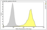
Evaluated by Flow Cytometry in human PBMCs.
Flow Cytometry Analysis: 0.2 µL of this antibody detected CD47 surface expression on the gated lymphocytes population among one million human PBMCs.
Usage Statement
Unless otherwise stated in our catalog or other company documentation accompanying the product(s), our products are intended for research use only and are not to be used for any other purpose, which includes but is not limited to, unauthorized commercial uses, in vitro diagnostic uses, ex vivo or in vivo therapeutic uses or any type of consumption or application to humans or animals.