Wenn Sie das Fenster schließen, wird Ihre Konfiguration nicht gespeichert, es sei denn, Sie haben Ihren Artikel in die Bestellung aufgenommen oder zu Ihren Favoriten hinzugefügt.
Klicken Sie auf OK, um das MILLIPLEX® MAP-Tool zu schließen oder auf Abbrechen, um zu Ihrer Auswahl zurückzukehren.
Wählen Sie konfigurierbare Panels & Premixed-Kits - ODER - Kits für die zelluläre Signaltransduktion & MAPmates™
Konfigurieren Sie Ihre MILLIPLEX® MAP-Kits und lassen sich den Preis anzeigen.
Konfigurierbare Panels & Premixed-Kits
Unser breites Angebot enthält Multiplex-Panels, für die Sie die Analyten auswählen können, die am besten für Ihre Anwendung geeignet sind. Unter einem separaten Register können Sie das Premixed-Cytokin-Format oder ein Singleplex-Kit wählen.
Kits für die zelluläre Signaltransduktion & MAPmates™
Wählen Sie gebrauchsfertige Kits zur Erforschung gesamter Signalwege oder Prozesse. Oder konfigurieren Sie Ihre eigenen Kits mit Singleplex MAPmates™.
Die folgenden MAPmates™ sollten nicht zusammen analysiert werden: -MAPmates™, die einen unterschiedlichen Assaypuffer erfordern. -Phosphospezifische und MAPmate™ Gesamtkombinationen wie Gesamt-GSK3β und Gesamt-GSK3β (Ser 9). -PanTyr und locusspezifische MAPmates™, z.B. Phospho-EGF-Rezeptor und Phospho-STAT1 (Tyr701). -Mehr als 1 Phospho-MAPmate™ für ein einziges Target (Akt, STAT3). -GAPDH und β-Tubulin können nicht mit Kits oder MAPmates™, die panTyr enthalten, analysiert werden.
.
Bestellnummer
Bestellinformationen
St./Pkg.
Liste
Dieser Artikel wurde zu Ihren Favoriten hinzugefügt.
Wählen Sie bitte Spezies, Panelart, Kit oder Probenart
Um Ihr MILLIPLEX® MAP-Kit zu konfigurieren, wählen Sie zunächst eine Spezies, eine Panelart und/oder ein Kit.
Custom Premix Selecting "Custom Premix" option means that all of the beads you have chosen will be premixed in manufacturing before the kit is sent to you.
Catalogue Number
Ordering Description
Qty/Pack
List
Dieser Artikel wurde zu Ihren Favoriten hinzugefügt.
Spezies
Panelart
Gewähltes Kit
Menge
Bestellnummer
Bestellinformationen
St./Pkg.
Listenpreis
96-Well Plate
Menge
Bestellnummer
Bestellinformationen
St./Pkg.
Listenpreis
Weitere Reagenzien hinzufügen (MAPmates erfordern die Verwendung eines Puffer- und Detektionskits)
Menge
Bestellnummer
Bestellinformationen
St./Pkg.
Listenpreis
48-602MAG
Buffer Detection Kit for Magnetic Beads
1 Kit
Platzsparende Option Kunden, die mehrere Kits kaufen, können ihre Multiplex-Assaykomponenten in Kunststoffbeuteln anstelle von Packungen erhalten, um eine kompaktere Lagerung zu ermöglichen.
Dieser Artikel wurde zu Ihren Favoriten hinzugefügt.
Das Produkt wurde in Ihre Bestellung aufgenommen
Sie können nun ein weiteres Kit konfigurieren, ein Premixed-Kit wählen, zur Kasse gehen oder das Bestell-Tool schließen.
SCC148
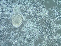
Sigma-AldrichMB49 Mouse Bladder Carcinoma Cell Line
MB49 mouse urothelial carcinoma cell line is widely used as an in vitro and in vivo model of bladder cancer.
More>>MB49 mouse urothelial carcinoma cell line is widely used as an in vitro and in vivo model of bladder cancer. Less<<
MB49 Mouse Bladder Carcinoma Cell Line: SDB (Sicherheitsdatenblätter), Analysenzertifikate und Qualitätszertifikate, Dossiers, Broschüren und andere verfügbare Dokumente.
α-Naphthyl Acid Phosphate, Monosodium Salt - CAS 81012-89-7 - Calbiochem
Übersicht
Replacement Information
Key Spec Table
Key Applications
CULT
Description
Catalogue Number
SCC148
Description
MB49 Mouse Bladder Carcinoma Cell Line
Overview
MB49 cells are derived from C57BL/Icrf-a’ mouse bladder epithelial cells that were transformed by a single 24-hour treatment with the chemical carcinogen 7, 12-dimethylbenz[a]anthracene (DMBA) on the second day of a long term primary culture (1). Transformed cells transplanted into syngeneic mice were shown to generate carcinomas (1). While of male origin, karyotype analyses indicate the loss of the Y chromosome in 100% of the cells analyzed (2). This abnormality is a frequent early event in human bladder cancer.
A recent study indicates that MB49 cells recapitulate key features of sex differences in bladder tumor growth (3). MB49 implantation in mice resulted in significantly larger tumors in males than females. In the presence of dihydrotestosterone, MB49 cells exhibited enhanced proliferation in a dose-dependent manner. In contrast, MB49 cells were unresponsive to the pregnancy hormone, human chorionic gonadotrophin (hCG) (3).
MB49 cells exhibit low to no expression of MHC-Class I and II molecules (4). However, upon exposure to IFN-, the expressions of MHC Class I and II are significantly upregulated (4).
References: 1. Summerhayes IC, Franks LM (1979) Effects of donor age on neoplastic transformation of adult mouse bladder epithelium in vitro. J Natl Cancer Inst 62(4): 1017 - 1023. 2. Fabris VT, Lodilinsky C, Pampena MB, Belgorosky D, Lanari C, Eiján AM (2012) Cytogenetic characterization of the murine bladder cancer model MB49 and the derived invasive line MB49-I. Cancer Genet 205(4): 168 - 76. 3. White-Gilbertson S, Davis M, Voelkel-Johnson C, Kasman LM (2016) Sex differences in the MB49 syngeneic, murine model of bladder cancer. Bladder (San Franc) 3(1): PMID: 26998503. 4. Lattime EC, Gomelia LG, McCue PA (1992) Murine bladder carcinoma cells present antigen to BCG-specific CD4+ T-cells. Cancer Res 52(15): 4286 - 90.
MB49 mouse urothelial carcinoma cell line is widely used as an in vitro and in vivo model of bladder cancer.
Key Applications
Cell Culture
Application Notes
This product is intended for sale and sold solely to academic institutions for internal academic research use per the terms of the “Academic Use Agreement” as detailed in the product documentation. For information regarding any other use, please contact licensing@emdmillipore.com.
Biological Information
Cell Line Type
Cancer Cells
Physicochemical Information
Dimensions
Materials Information
Toxicological Information
Safety Information according to GHS
Safety Information
Product Usage Statements
Quality Assurance
• Each vial contains ≥ 1X10⁶ viable cells. • Cells are tested negative for infectious diseases by a Mouse Essential CLEAR panel by Charles River Animal Diagnostic Services. • Cells are verified to be of mouse origin and negative for inter-species contamination from rat, chinese hamster, Golden Syrian hamster, human and non-human primate (NHP) as assessed by a Contamination CLEAR panel by Charles River Animal Diagnostic Services. • Cells are negative for mycoplasma contamination
Usage Statement
Unless otherwise stated in our catalog or other company documentation accompanying the product(s), our products are intended for research use only and are not to be used for any other purpose, which includes but is not limited to, unauthorized commercial uses, in vitro diagnostic uses, ex vivo or in vivo therapeutic uses or any type of consumption or application to humans or animals.