Le fait de fermer ne sauvegardera pas votre configuration à moins que vous n'ajoutiez l'article à votre Panier d'achat ou à vos Favoris.
Cliquer sur OK pour fermer l'outil MILLIPLEX® MAP ou sur Annuler pour retourner à votre sélection.
Choisissez des Panels configurables & des Kits préconfigurés - OU - des MAPmate™ de signalisation cellulaire
Concevez vos kits MILLIPLEX® MAP et obtenez leur prix.
Panels configurables & Kits préconfigurés
Notre large gamme est constituée de panels multiplex qui vous permettent de choisir, au sein d'un panel, les analytes qui répondent le mieux à vos besoins. Sur un autre onglet, vous pouvez choisir un format cytokine préconfiguré ou un kit Simplex.
Kits de signalisation cellulaire & MAPmate™
Choisissez des kits préconfigurés qui permettent d'explorer l'ensemble des voies ou des processus. Ou concevez vos propres kits en choisissant des Simplex MAPmate™ et en suivant les instructions fournies.
Les MAPmate™ suivants ne peuvent pas être utilisés ensemble : -des MAPmate™ qui nécessitent des tampons différents -des paires de MAPmate™ totaux et phospho-spécifiques, par ex. GSK3β total et GSK3β (Ser 9) -des MAPmate™ PanTyr et spécifiques d'un site, par ex. Récepteur Phospho-EGF et phospho-STAT1 (Tyr701) -Plus d'un phospho-MAPmate™ pour une seule cible (Akt, STAT3). -GAPDH et β-Tubuline ne peuvent pas être utilisés avec les kits ou les MAPmate™ contenant panTyr.
.
Référence
Guide d'achat
Qté
Liste
Cet article a été ajouté à vos favoris.
Sélectionner une espèce, un type de panel, un kit ou un type d'échantillon
Pour commencer à concevoir votre kit MILLIPLEX® MAP, sélectionnez une espèce, un type de panel ou un kit d'intérêt.
Custom Premix Selecting "Custom Premix" option means that all of the beads you have chosen will be premixed in manufacturing before the kit is sent to you.
Catalogue Number
Ordering Description
Qty/Pack
List
Cet article a été ajouté à vos favoris.
Espèce
Type de panel
Kit sélectionné
Qté
Référence
Guide d'achat
Qté
Prix tarif
96-Well Plate
Qté
Référence
Guide d'achat
Qté
Prix tarif
Ajouter des réactifs supplémentaires (Un kit "Buffer and Detection Kit" est nécessaire pour une utilisation avec les MAPmate™)
Qté
Référence
Guide d'achat
Qté
Prix tarif
48-602MAG
Buffer Detection Kit for Magnetic Beads
1 Kit
Option de gain de place Nos clients qui commandent plusieurs kits peuvent choisir d'économiser de l'espace de stockage en éliminant l'emballage de chaque kit et de recevoir les composants de leur essai multiplex conditionnés sous poches en plastique pour un stockage plus compact.
Cet article a été ajouté à vos favoris.
Ce produit a été ajouté à votre panier.
Vous pouvez maintenant concevoir un autre kit personnalisé, choisir un kit pré-configuré, régler vos achats ou fermer l'outil de commande.
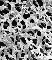
Filtres membranes Durapore®
Durapore® membranes made with Polyvinylidene fluoride (PVDF) provide high flow rates and throughput, low extractables and broad chemical compatibility.Plus
Durapore® membranes made with Polyvinylidene fluoride (PVDF) provide high flow rates and throughput, low extractables and broad chemical compatibility. Moins
Colony filtration blot: a new screening method for soluble protein expression in Escherichia coli Tobias Cornvik, Sue-Li Dahlroth, Audur Magnusdottir, Maria Dolores Herman, Rosemarie Knaust, Monica Ekberg & Pär Nordlund Nature Methods, 2005 Jul:2(7):507-509
2004
A functional study on polymorphism of the ATP-binding Cassette transpoter ABCG2: Toshihisa Ishikawa,Biochem.J, 373, 767-774, 2003, internal ref ABC-02 Biochem.J, 373, 767-774, 2003, internal ref ABC-02
2003
Comparison of microporous membrane morphologies using confocal scanning laser microscopy Charcosset C (reprint), et al. Journal of membrane science. 2000. v168, n2, p53-62
1999
ATP/Mg2+-dependent Cardiac Transport system for Flutathione S-Conjugates Toshihisa Ishikawa,J.Biological Chemistry, 264:29, 17343-17348, 1989, internal ref ABC-01 J.Biological Chemistry, 264:29, 17343-17348, 1989, internal ref ABC-01
1988
FAQ
Question
Réponse
How do I tell the membrane from the separator?
The blue paper is the separator.
How do I assemble a diffusion chamber containing tissue for implantation?
Wet a single membrane filter (either GSWP 013 00 or HAWP 013 00) with Milli-Q water and blot it to absorbent paper to remove excess water.
Glue the filter to one side of a diffusion chamber ring without hole (catalog number PR00 014 00), using MF cement (catalog number XX70 000 00), and let dry.
Sterilize this assembly, along with another membrane filter of the same type, by exposure to ethylene oxide gas or ultraviolet light.
Moisten the half-completed chamber and the separate sterile filter with a sterile physiological solution, and aseptically place the tissue to be studied in the half-completed chamber assembly.
Glue the cover filter in place using sterile technique.
What are the dimensions and width of the grids on the Millipore 47mm gridded membranes?
The grids are 3.08mm x 3.08mm square. A grid line is 44um in width and there is are average of 169 gridded squares per filter. Please note however that the actual filtration area and therefore the total number of squares in that area is dependent on the filter holder. For filter holders with 9.6sq.cm of filtration area there are 100 squares available. For filter holders with 13.8 sq.cm of filtration area the number of available grids are approximately 140.
How do I assemble a diffusion chamber containing suspension cells for implantation?
Wet two membrane filters (either GSWP 013 00 or HAWP 013 00) with Milli-Q water and blot them to absorbent paper to remove excess water. Glue a moist filter to each side of the diffusion chamber ring with hole (catalog number PR00 014 01) using MF cement (catalog number XX70 000 00). Sterilize the dried diffusion chamber with ethylene oxide gas or ultraviolet light. After sterilization, inject the cells through the hole in the plastic ring, and plug the hole with the nylon thread which comes with the rings (catalog number PR00 000 00 if ordered separately).
Can Millipore filters remove mycoplasma from my solution?
Mycoplasma can be removed from solutions by using a 0.1 um pore size filter. For mycoplasma reduction the 0.1 um Durapore filter can be used. Its Millipore code is VV. A 0.1 um Express membrane can also be used for removal of mycoplasma. Its membrane code is VP. These membranes are available in a number of fabricated fiter devices. For help determining which device would be best for your solution, call Millipore Technical Services.
Are the pore sizes of your Durapore membranes uniform or asymmetric?
The surfaces of the Durapore membrane are symmetric, however, the actual pores are not straight-through holes but rather follow a torturous path.
Which membrane should I use to filter my solution?
For general filtration use MCE. When you are looking for the lowest protein binding membrane use Durapore. For speed, and when filtering serum solutions use Millipore Express.
I would like to use the Durapore membrane discs and am interested in knowing what is meant by average pore size?
Since the size and uniformity of the Durapore membrane is accurately controlled during manufacturing, it is possible to rate membranes as absolute filters based on the largest particle that can pass through the membrane. It is then possible to confirm the pore size rating of the membrane filter using a non-destructive integrity test referred to as the bubble point test. This test is based on the fact that liquid is held in the membrane pores by surface tension and that the minimum pressure required to force liquid from the capillary structure is directly related to the capillary diameter.
Which side of the membrane should I use, the shiny or dull side?
Most researchers may not even notice that there is a "sidedness" to filters, and, for most applications, orientation will not affect filter performance. However, membranes do have a slightly asymmetric pore structure: the shiny side of the membrane is the "tighter" side. In some applications, you can take advantage of this difference by selecting a specific filter orientation. ultrafiltration membranes should always be used shiny side up, regardless of application for drop dialysis ( a buffer exchange technique in which a few drops of DNA or protein are placed in a 0.05 or 0.025 um filter and floated on a buffer solution), apply the sample to the shiny side of the filter and float the filter dull side to the buffer. This measure will enhance buffer exchange and discourage sample loss. The Millipore Express and Express Plus membranees are also sided - these membranes should be used shiny side facing down.
I have a dilute protein solution. I want to make sure I do not lose any of my protein. Which filter should I use?
You should use any device that contains a Durapore filter. Durapore is the lowest protein-binding filter available. This filter will give the best yield when filtering a dilute protein solution.


)