Astroblastoma: beside being a tumor entity, an occasional phenotype of astrocytic gliomas?
Mellai, M; Piazzi, A; Casalone, C; Grifoni, S; Melcarne, A; Annovazzi, L; Cassoni, P; Denysenko, T; Valentini, MC; Cistaro, A; Schiffer, D
OncoTargets and therapy
8
451-60
2015
Show Abstract
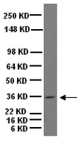
The diagnosis of astroblastoma is based on a typical histological aspect with perivascular distribution of cells sending cytoplasmic extensions to the vessels and vascular hyalinization. These criteria are useful for standardizing the identification of the tumor, but, in spite of this, there are discrepancies in the literature concerning the age distribution and the benign or malignant nature of the tumor. Three cases are discussed in this study: Case 1 was a typical high-grade astroblastoma; Case 2 was an oligodendroglioma at the first intervention and an oligoastrocytoma at the second intervention with typical perivascular arrangements in the astrocytic component; Case 3 was a gemistocytic glioma with malignant features and typical perivascular arrangements. Genetic analysis showed genetic alterations that are typical of gliomas of all malignancy grades. Using the neurosphere assay, neurospheres and adherent cells were found to have developed in Case 1, while adherent cells only developed in Case 2, in line with the stemness potential of the tumors. The cases are discussed in relation to their diagnostic assessment as astroblastoma, and it is hypothesized that the typical perivascular distribution of cells may not indicate a separate and unique tumor entity, but may be a peculiarity that can be acquired by astrocytic gliomas when an unknown cause from the tumor microenvironment influences the relationship between vessels and tumor cells. | Immunofluorescence | | 25737639
 |
Cell lineage identification and stem cell culture in a porcine model for the study of intestinal epithelial regeneration.
Gonzalez, LM; Williamson, I; Piedrahita, JA; Blikslager, AT; Magness, ST
PloS one
8
e66465
2013
Show Abstract
Significant advances in intestinal stem cell biology have been made in murine models; however, anatomical and physiological differences between mice and humans limit mice as a translational model for stem cell based research. The pig has been an effective translational model, and represents a candidate species to study intestinal epithelial stem cell (IESC) driven regeneration. The lack of validated reagents and epithelial culture methods is an obstacle to investigating IESC driven regeneration in a pig model. In this study, antibodies against Epithelial Adhesion Molecule 1 (EpCAM) and Villin marked cells of epithelial origin. Antibodies against Proliferative Cell Nuclear Antigen (PCNA), Minichromosome Maintenance Complex 2 (MCM2), Bromodeoxyuridine (BrdU) and phosphorylated Histone H3 (pH3) distinguished proliferating cells at various stages of the cell cycle. SOX9, localized to the stem/progenitor cells zone, while HOPX was restricted to the +4/'reserve' stem cell zone. Immunostaining also identified major differentiated lineages. Goblet cells were identified by Mucin 2 (MUC2); enteroendocrine cells by Chromogranin A (CGA), Gastrin and Somatostatin; and absorptive enterocytes by carbonic anhydrase II (CAII) and sucrase isomaltase (SIM). Transmission electron microscopy demonstrated morphologic and sub-cellular characteristics of stem cell and differentiated intestinal epithelial cell types. Quantitative PCR gene expression analysis enabled identification of stem/progenitor cells, post mitotic cell lineages, and important growth and differentiation pathways. Additionally, a method for long-term culture of porcine crypts was developed. Biomarker characterization and development of IESC culture in the porcine model represents a foundation for translational studies of IESC-driven regeneration of the intestinal epithelium in physiology and disease. | | | 23840480
|
Musashi1-CreER(T2) : a new cre line for conditional mutagenesis in neural stem cells.
Takeda, H; Koso, H; Tessarollo, L; Copeland, NG; Jenkins, NA
Genesis (New York, N.Y. : 2000)
51
128-34
2013
Show Abstract
The RNA-binding protein Musashi1 (Msi1) is one of two mammalian homologues of Drosophila Musashi, which is required for the asymmetric cell division of sensory organ precursor cells. In the mouse central nervous system (CNS), Msi1 is preferentially expressed in mitotically active progenitor cells in the ventricular zone (VZ) of the neural tube during embryonic development and in the subventricular zone (SVZ) of the postnatal brain. Previous studies showed that cells in the SVZ can contribute to long-term neurogenesis in the olfactory bulb (OB), but it remains unclear whether Msi1-expressing cells have self-renewing potential and can contribute to neurogenesis in the adult. Here, we describe the generation of Msi1-CreER(T2) knock-in mice and show by cell lineage tracing that Msi1-CreER(T2) -expressing cells mark neural stem cells (NSCs) in both the embryonic and adult brain. Msi1-CreER(T2) mice thus represent a new tool in our arsenal for genetically manipulating NSCs, which will be essential for understanding the molecular mechanisms underlying neural development. | | | 23132814
|
Progenitor-like traits contribute to patient survival and prognosis in oligodendroglial tumors.
Ng, FS; Toh, TB; Ting, EH; Koh, GR; Sandanaraj, E; Phong, M; Wong, SS; Leong, SH; Kon, OL; Tucker-Kellogg, G; Ng, WH; Ng, I; Tang, C; Ang, BT
Clinical cancer research : an official journal of the American Association for Cancer Research
18
4122-35
2012
Show Abstract
Patient-derived glioma-propagating cells (GPC) contain karyotypic and gene expression profiles that are found in the primary tumor. However, their clinical relevance is unclear. We ask whether GPCs contribute to disease progression and survival outcome in patients with glioma by analyzing gene expression profiles.We tapped into public sources of GPC gene expression data and derived a gene signature distinguishing oligodendroglial from glioblastoma multiforme (GBM) GPCs. By adapting a method in glioma biology, the Connectivity Map, we interrogated its strength of association in public clinical databases. We validated the top-ranking signaling pathways Wnt, Notch, and TGFβ, in GPCs and primary tumor specimens.We observed that patients with better prognosis correlated with oligodendroglial GPC features and lower tumor grade, and this was independent of the current clinical indicator, 1p/19q status. Patients with better prognosis had proneural tumors whereas the poorly surviving cohort had mesenchymal tumors. In addition, oligodendroglial GPCs were more sensitive to Wnt and Notch inhibition whereas GBM GPCs responded to TGFβR1 inhibition.We provide evidence that GPCs are clinically relevant. In addition, the more favorable prognosis of oligodendroglial tumors over GBM could be recapitulated transcriptomically at the GPC level, underscoring the relevance of this cellular model. Our gene signature detects molecular heterogeneity in oligodendroglial tumors that cannot be accounted for by the 1p/19q status alone, indicating that stem-like traits contribute to clinical status. Collectively, these data highlight the limitation of morphology-based histologic analyses in tumor classification, consequently impacting on treatment decisions. | | | 22675171
|
IKKβ/NF-κB disrupts adult hypothalamic neural stem cells to mediate a neurodegenerative mechanism of dietary obesity and pre-diabetes.
Li, J; Tang, Y; Cai, D
Nature cell biology
14
999-1012
2012
Show Abstract
Adult neural stem cells (NSCs) are known to exist in a few regions of the brain; however, the entity and physiological/disease relevance of adult hypothalamic NSCs (htNSCs) remain unclear. This work shows that adult htNSCs are multipotent and predominantly present in the mediobasal hypothalamus of adult mice. Chronic high-fat-diet feeding led to not only depletion but also neurogenic impairment of htNSCs associated with IKKβ/NF-κB activation. In vitro htNSC models demonstrated that their survival and neurogenesis markedly decreased on IKKβ/NF-κB activation but increased on IKKβ/NF-κB inhibition, mechanistically mediated by IKKβ/NF-κB-controlled apoptosis and Notch signalling. Mouse studies revealed that htNSC-specific IKKβ/NF-κB activation led to depletion and impaired neuronal differentiation of htNSCs, and ultimately the development of obesity and pre-diabetes. In conclusion, adult htNSCs are important for the central regulation of metabolic physiology, and IKKβ/NF-κB-mediated impairment of adult htNSCs is a critical neurodegenerative mechanism for obesity and related diabetes. | Immunofluorescence | | 22940906
|
The C-terminus of Apc does not influence intestinal adenoma development or progression.
Annabelle Lewis,Hayley Davis,Maesha Deheragoda,Patrick Pollard,Emma Nye,Rosemary Jeffery,Stefania Segditsas,Philip East,Richard Poulsom,Gordon Stamp,Nicholas Wright,Ian Tomlinson
The Journal of pathology
226
2012
Show Abstract
Adenomatous polyposis coli (APC ) mutations are found in most colorectal tumours. These mutations are almost always protein-truncating, deleting both central domains that regulate Wnt signalling and C-terminal domains that interact with the cytoskeleton. The importance of Wnt dysregulation for colorectal tumourigenesis is well characterized. It is, however, unclear whether loss of C-terminal functions contributes to tumourigenesis, although this protein region has been implicated in cellular processes--including polarity, migration, mitosis, and chromosomal instability (CIN)—that have been postulated as critical for the development and progression of intestinal tumours. Since almost all APC mutations in human patients disrupt both central and C-terminal regions, we created a mouse model to test the role of the C-terminus of APC in intestinal tumourigenesis. This mouse (Apc(ΔSAMP)) carries an internal deletion within Apc that dysregulates Wnt by removing the beta-catenin-binding and SAMP repeats, but leaves the C-terminus intact. We compared Apc(ΔSAMP) mice with Apc(1322T) animals. The latter allele represented the most commonly found human APC mutation and was identical to Apc(ΔSAMP) except for absence of the entire C-terminus. Apc(ΔSAMP) mice developed numerous intestinal adenomas indistinguishable in number, location, and dysplasia from those seen in Apc(1322T) mice. No carcinomas were found in Apc(ΔSAMP) or Apc(1322T) animals. While similar disruption of the Wnt signalling pathway was observed in tumours from both mice, no evidence of differential C-terminus functions (such as cell migration, CIN, or localization of APC and EB1) was seen. We conclude that the C-terminus of APC does not influence intestinal adenoma development or progression. | | | 22009253
|
Functional neural stem cell isolation from brains of adult mutant SOD1 (SOD1(G93A)) transgenic amyotrophic lateral sclerosis (ALS) mice.
Lee JC, Jin Y, Jin J, Kang BG, Nam DH, Joo KM, Cha CI
Neurol Res
33
33-7. Epub 2010 Aug 31.
2011
Show Abstract
OBJECTIVES: The aim of present study is to investigate more functional neural stem cells (NSCs) could be isolated from brains with amyotrophic lateral sclerosis (ALS) and expanded in vitro, based on previous reports demonstrating de novo neurogenesis is enhanced to replace degenerating neural tissue.METHODS: Thirteen- or eighteen-week-old mutant human Cu/Zn superoxide dismutase (SOD1(G93A)) transgenic ALS and wild-type SOD1 transgenic control mice were utilized. Changes in numbers of NSCs in the dentate gyrus were analyzed by immunohistochemistry against nestin and CD133. NSCs were primarily cultured from hippocampus of ALS or control mice. Expression of NSC markers, in vitro expansion capacity, and differentiating potential were compared.RESULTS: Hippocampus of 13-week-old pre-symptomatic ALS mice harbor more cells that can be propagated for more than 12 passages in vitro, compared with same age control mice. Primarily-cultured cells formed neurospheres in the NSC culture medium, expressed NSC markers, and differentiated into cells with differentiated neural cell characteristics in the differentiation condition confirming that they are NSCs. In contrast, long-term expansible NSCs could not be derived from brains of 18-week-old symptomatic ALS mice with the same experimental techniques, although they had comparable nestin-immunoreactive cells in the dentate gyrus.DISCUSSION: These results would suggest that increased neuroregeneration in early phase of ALS could be translated to regenerative approaches; however, long-term exposure to ALS microenvironments could abolish functional capacities of NSCs. | | | 20810028
|
Changes in Musashi-1 subcellular localization correlate with cell cycle exit during postnatal retinal development.
P E B Nickerson,T Myers,D B Clarke,R L Chow
Experimental eye research
92
2011
Show Abstract
RNA-binding proteins, and in particular, the Musashi genes, function as essential regulators of progenitor functioning in both the developing and adult organism. In this report, we characterize the differential subcellular distribution of Musashi-1 in cells engaged in either proliferating or differentiating contexts in the developing mouse retina, and in cultured Müller glia. During retinal cell differentiation, Musashi-1 immunoreactivity shifts from exclusively cytoplasmic in retinal progenitor cells, to predominantly nuclear localization in differentiating neurons. This nuclear shift is transient, with localization in the adult retina becoming predominantly perinuclear and cytoplasmic in Müller glia and photoreceptors. A correlation between cell cycle progression and subcellular distribution of Musashi-1 is observed in passageable, adult Müller glial cells in vitro. Furthermore, treatment of Müller cultures with neuron-promoting differentiation media induces asymmetric cytoplasmic Musashi-1 immunoreactivity in dividing daughter cells. The observed shifts in subcellular Musashi-1 localization are consistent with contrasting roles for Musashi-1 during cell proliferation and differentiation. These data provide evidence that nuclear, and cytoplasmic sequestering of Musashi-1 in retinal cells is context-specific, and may contribute to downstream functioning of Musashi-1. | | | 21320487
|
A novel population of myeloid cells responding to coxsackievirus infection assists in the dissemination of virus within the neonatal CNS.
Tabor-Godwin, JM; Ruller, CM; Bagalso, N; An, N; Pagarigan, RR; Harkins, S; Gilbert, PE; Kiosses, WB; Gude, NA; Cornell, CT; Doran, KS; Sussman, MA; Whitton, JL; Feuer, R
The Journal of neuroscience : the official journal of the Society for Neuroscience
30
8676-91
2010
Show Abstract
Enterovirus infection in newborn infants is a significant cause of aseptic meningitis and encephalitis. Using a neonatal mouse model, we previously determined that coxsackievirus B3 (CVB3) preferentially targets proliferating neural stem cells located in the subventricular zone within 24 h after infection. At later time points, immature neuroblasts, and eventually mature neurons, were infected as determined by expression of high levels of viral protein. Here, we show that blood-derived Mac3(+) mononuclear cells were rapidly recruited to the CNS within 12 h after intracranial infection with CVB3. These cells displayed a myeloid-like morphology, were of a peripheral origin based on green fluorescent protein (GFP)-tagged adoptive cell transplant examination, and were highly susceptible to CVB3 infection during their migration into the CNS. Serial immunofluorescence images suggested that the myeloid cells enter the CNS via the choroid plexus, and that they may be infected during their extravasation and passage through the choroid plexus epithelium; these infected myeloid cells ultimately penetrate into the parenchyma of the brain. Before their migration through the ependymal cell layer, a subset of these infected myeloid cells expressed detectable levels of nestin, a marker for neural stem and progenitor cells. As these nestin(+) myeloid cells infected with CVB3 migrated through the ependymal cell layer, they revealed distinct morphological characteristics typical of type B neural stem cells. The recruitment of these novel myeloid cells may be specifically set in motion by the induction of a unique chemokine profile in the CNS induced very early after CVB3 infection, which includes upregulation of CCL12. We propose that intracranial CVB3 infection may lead to the recruitment of nestin(+) myeloid cells into the CNS which might represent an intrinsic host CNS repair response. In turn, the proliferative and metabolic status of recruited myeloid cells may render them attractive targets for CVB3 infection. Moreover, the migratory ability of these myeloid cells may point to a productive method of virus dissemination within the CNS. Full Text Article | Immunofluorescence | | 20573913
|
Musashi-2 regulates normal hematopoiesis and promotes aggressive myeloid leukemia.
Michael G Kharas,Christopher J Lengner,Fatima Al-Shahrour,Lars Bullinger,Brian Ball,Samir Zaidi,Kelly Morgan,Winnie Tam,Mahnaz Paktinat,Rachel Okabe,Maricel Gozo,William Einhorn,Steven W Lane,Claudia Scholl,Stefan Fröhling,Mark Fleming,Benjamin L Ebert,D Gary Gilliland,Rudolf Jaenisch,George Q Daley
Nature medicine
16
2010
Show Abstract
RNA-binding proteins of the Musashi (Msi) family are expressed in stem cell compartments and in aggressive tumors, but they have not yet been widely explored in the blood. Here we demonstrate that Msi2 is the predominant form expressed in hematopoietic stem cells (HSCs), and its knockdown leads to reduced engraftment and depletion of HSCs in vivo. Overexpression of human MSI2 in a mouse model increases HSC cell cycle progression and cooperates with the chronic myeloid leukemia-associated BCR-ABL1 oncoprotein to induce an aggressive leukemia. MSI2 is overexpressed in human myeloid leukemia cell lines, and its depletion leads to decreased proliferation and increased apoptosis. Expression levels in human myeloid leukemia directly correlate with decreased survival in patients with the disease, thereby defining MSI2 expression as a new prognostic marker and as a new target for therapy in acute myeloid leukemia (AML). Full Text Article | | | 20616797
|