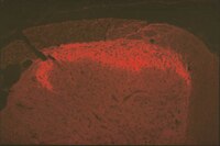
Spinal changes of a newly isolated neuropeptide endomorphin-2 concomitant with vincristine-induced allodynia.
Yang, Y; Zhang, YG; Lin, GA; Xie, HQ; Pan, HT; Huang, BQ; Liu, JD; Liu, H; Zhang, N; Li, L; Chen, JH
PloS one
9
e89583
2014
Show Abstract
Chemotherapy-induced neuropathic pain (CNP) is the major dose-limiting factor in cancer chemotherapy. However, the neural mechanisms underlying CNP remain unclear. There is increasing evidence implicating the involvement of spinal endomorphin-2 (EM2) in neuropathic pain. In this study, we used a vincristine-evoked rat CNP model displaying mechanical allodynia and central sensitization, and observed a significant decrease in the expression of spinal EM2 in CNP. Also, while intrathecal administration of exogenous EM2 attenuated allodynia and central sensitization, the mu-opioid receptor antagonist β-funaltrexamine facilitated these events. We found that the reduction in spinal EM2 was mediated by increased activity of dipeptidylpeptidase IV, possibly as a consequence of chemotherapy-induced oxidative stress. Taken together, our findings suggest that a decrease in spinal EM2 expression causes the loss of endogenous analgesia and leads to enhanced pain sensation in CNP. | Immunohistochemistry | 24586889
 |
A major external source of cholinergic innervation of the striatum and nucleus accumbens originates in the brainstem.
Dautan, D; Huerta-Ocampo, I; Witten, IB; Deisseroth, K; Bolam, JP; Gerdjikov, T; Mena-Segovia, J
The Journal of neuroscience : the official journal of the Society for Neuroscience
34
4509-18
2014
Show Abstract
Cholinergic transmission in the striatal complex is critical for the modulation of the activity of local microcircuits and dopamine release. Release of acetylcholine has been considered to originate exclusively from a subtype of striatal interneuron that provides widespread innervation of the striatum. Cholinergic neurons of the pedunculopontine (PPN) and laterodorsal tegmental (LDT) nuclei indirectly influence the activity of the dorsal striatum and nucleus accumbens through their innervation of dopamine and thalamic neurons, which in turn converge at the same striatal levels. Here we show that cholinergic neurons in the brainstem also provide a direct innervation of the striatal complex. By the expression of fluorescent proteins in choline acetyltransferase (ChAT)::Cre(+) transgenic rats, we selectively labeled cholinergic neurons in the rostral PPN, caudal PPN, and LDT. We show that cholinergic neurons topographically innervate wide areas of the striatal complex: rostral PPN preferentially innervates the dorsolateral striatum, and LDT preferentially innervates the medial striatum and nucleus accumbens core in which they principally form asymmetric synapses. Retrograde labeling combined with immunohistochemistry in wild-type rats confirmed the topography and cholinergic nature of the projection. Furthermore, transynaptic gene activation and conventional double retrograde labeling suggest that LDT neurons that innervate the nucleus accumbens also send collaterals to the thalamus and the dopaminergic midbrain, thus providing both direct and indirect projections, to the striatal complex. The differential activity of cholinergic interneurons and cholinergic neurons of the brainstem during reward-related paradigms suggest that the two systems play different but complementary roles in the processing of information in the striatum. | Immunohistochemistry | 24671996
 |
Synaptic connections between endomorphin 2-immunoreactive terminals and μ-opioid receptor-expressing neurons in the sacral parasympathetic nucleus of the rat.
Dou, XL; Qin, RL; Qu, J; Liao, YH; Lu, Yc; Zhang, T; Shao, C; Li, YQ
PloS one
8
e62028
2013
Show Abstract
The urinary bladder is innervated by parasympathetic preganglionic neurons (PPNs) that express μ-opioid receptors (MOR) in the sacral parasympathetic nucleus (SPN) at lumbosacral segments L6-S1. The SPN also contains endomorphin 2 (EM2)-immunoreactive (IR) fibers and terminals. EM2 is the endogenous ligand of MOR. In the present study, retrograde tract-tracing with cholera toxin subunit b (CTb) or wheat germ agglutinin-conjugated horseradish peroxidase (WGA-HRP) via the pelvic nerve combined with immunohistochemical staining for EM2 and MOR to identify PPNs within the SPN as well as synaptic connections between the EM2-IR terminals and MOR-expressing PPNs in the SPN of the rat. After CTb was injected into the pelvic nerve, CTb retrogradely labeled neurons were almost exclusively located in the lateral part of the intermediolateral gray matter at L6-S1 of the lumbosacral spinal cord. All of the them also expressed MOR. EM2-IR terminals formed symmetric synapses with MOR-IR, WGA-HRP-labeled and WGA-HRP/MOR double-labeled neuronal cell bodies and dendrites within the SPN. These results provided morphological evidence that EM2-containing axon terminals formed symmetric synapses with MOR-expressing PPNs in the SPN. The present results also show that EM2 and MOR might be involved in both the homeostatic control and information transmission of micturition. | Immunohistochemistry | 23671582
 |
Enhanced dendritic availability of μ-opioid receptors in inhibitory neurons of the extended amygdala in Mice deficient in the corticotropin-releasing factor-1 receptor.
Jaferi A, Zhou P, Pickel VM
Synapse (New York, NY)
65
8-20. doi
2011
Show Abstract
Activation of the corticotropin-releasing factor-1 (CRF-1) receptor in the anterolateral BNST (BSTal), a key subdivision of the extended amygdala, elicits opiate-seeking behavior exacerbated by stress. However, it is unknown whether the presence of CRF-1 affects expression of the μ-opioid receptor (μ-OR) in the many GABAergic BSTal neurons implicated in the stress response. We hypothesized that deletion of the CRF-1 receptor gene would alter the density and/or subcellular distribution of μ-ORs in GABAergic neurons of the BSTal. We used electron microscopy to quantitatively examine μ-OR immunogold and γ-aminobutyric acid (GABA) immunoperoxidase labeling in the BSTal of CRFr-1 knockout (KO) compared to wild-type (WT) mice. To assess regional specificity, we examined μ-OR distribution in dorsal striatum. The μ-ORs in each region were predominantly localized in dendrites, many of which were GABA-immunoreactive. Significantly, more cytoplasmic μ-OR gold particles per dendritic area were observed selectively in GABA-containing dendrites of the BSTal, but not of the dorsal striatum, in KO compared to WT mice. In both regions, however, significantly fewer GABA-immunoreactive axon terminals were present in KO compared to WT mice. Our results suggest that the absence of CRF-1 results in enhanced expression and/or dendritic trafficking of μ-ORs in inhibitory BSTal neurons. They also suggest that the expression of CRF-1 is a critical determinant of the availability of GABA in functionally diverse brain regions. These findings underscore the complex interplay between CRF, opioid, and GABA systems in limbic and striatal regions and have implications for the role of CRF-1 in influencing the pharmacological effects of opiates active at μ-ORs.Copyright © 2010 Wiley-Liss, Inc. | | 20506149
 |
Partial biopterin deficiency disturbs postnatal development of the dopaminergic system in the brain.
Homma, D; Sumi-Ichinose, C; Tokuoka, H; Ikemoto, K; Nomura, T; Kondo, K; Katoh, S; Ichinose, H
The Journal of biological chemistry
286
1445-52
2011
Show Abstract
Postnatal development of dopaminergic system is closely related to the development of psychomotor function. Tyrosine hydroxylase (TH) is the rate-limiting enzyme in the biosynthesis of dopamine and requires tetrahydrobiopterin (BH4) as a cofactor. To clarify the effect of partial BH4 deficiency on postnatal development of the dopaminergic system, we examined two lines of mutant mice lacking a BH4-biosynthesizing enzyme, including sepiapterin reductase knock-out (Spr(-/-)) mice and genetically rescued 6-pyruvoyltetrahydropterin synthase knock-out (DPS-Pts(-/-)) mice. We found that biopterin contents in the brains of these knock-out mice were moderately decreased from postnatal day 0 (P0) and remained constant up to P21. In contrast, the effects of BH4 deficiency on dopamine and TH protein levels were more manifested during the postnatal development. Both of dopamine and TH protein levels were greatly increased from P0 to P21 in wild-type mice but not in those mutant mice. Serotonin levels in those mutant mice were also severely suppressed after P7. Moreover, striatal TH immunoreactivity in Spr(-/-) mice showed a drop in the late developmental stage, when those mice exhibited hind-limb clasping behavior, a type of motor dysfunction. Our results demonstrate a critical role of biopterin in the augmentation of TH protein in the postnatal period. The developmental manifestation of psychomotor symptoms in BH4 deficiency might be attributable at least partially to high dependence of dopaminergic development on BH4 availability. | | 21062748
 |
Induction of Fos proteins in regions of the nucleus accumbens and ventrolateral striatum correlates with catalepsy and stereotypic behaviours induced by morphine.
Adam S Hamlin,Gavan P McNally,R Fred Westbrook,Peregrine B Osborne
Neuropharmacology
56
2009
Show Abstract
A history of intermittent exposures to drugs of abuse can cause long-term changes in acute behavioural responses to a subsequent drug exposure. In drug-naive rats, morphine can elicit intermittent cataleptic postures followed by sustained increases in locomotor activity. Chronic intermittent morphine treatment can reduce catalepsy and increase locomotor behaviour and stereotypy induced by morphine, even after prolonged periods of abstinence. The nucleus accumbens and limbic basal ganglia circuitry are implicated in the expression of various morphine-induced motor behaviours and catalepsy. We examined the effect of intermittent morphine exposure on the distribution of Fos proteins in the basal ganglia following a subsequent morphine challenge administered after a period of drug abstinence. We found that such exposures increased c-Fos induced by a morphine challenge in accumbens core regions that were immunoreactive for the micro-opioid receptor, and this correlated with the frequency of stereotypic behaviours displayed by the rats. We also found that a history of morphine exposures increased c-Fos in the ventrolateral striatum in response to a morphine challenge following 14 d but not 24 h of drug abstinence. In contrast, such a history induced acute Fras in the nucleus accumbens in response to a morphine challenge following 24 h but not 14 d of morphine abstinence. These data provide further confirmation that psychomotor sensitisation induced by repetitive morphine exposure involves long-term neuroadaptations in basal ganglia circuitry particularly at the level of the nucleus accumbens. | | 19705550
 |
Developmental changes of serotonin 4(a) receptor expression in the rat pre-Bötzinger complex.
Till Manzke,Stefan Preusse,Swen Hülsmann,Diethelm W Richter
The Journal of comparative neurology
506
2008
Show Abstract
Serotonin receptors (5-HTRs) are known to be involved in the regulation of breathing behavior and to mediate neurotrophic actions that exert a significant function in network formation during development. We studied neuronal 5-HT(4(a))R-immunoreactivity (-IR) at developmental ages from E14 to P10. Within the pre-Bötzinger complex (pre-BötC), a part of the respiratory network important for rhythmogenesis, 5-HT(4(a))R-IR was most extensive in rats at an age of E18. The 5-HT(4(a))-IR was found predominantly in the neuropil, whereas somatic staining was sporadic at late embryonic (E18-E20) stages. At birth, we observed a dramatic change to a predominantly somatic staining, and neuropil staining was greatly reduced and disappeared at an age of P4. In all developmental stages, 5-HT(4(a)) and mu-opioid receptors were strongly coexpressed in neurons of the pre-BötC, whereas 5-HT(4(a))R expression was absent in neurons within the dorsal horn. Nestin, a marker for CNS progenitor cells, was used to obtain information about the degree of pre-BötC differentiation. Nestin-positive cells did not appear within the pre-BötC before age E20. At E16, nestin-expressing cells were absent in the nucleus ambiguus (NA) and its ventral periphery. The number of nestin-positive cells increased after birth within and outside the pre-BötC, the majority of cells being glial. Coexpression of nestin and 5-HT(4(a))R was localized predominantly within the NA and appeared only sporadically within the pre-BötC. We conclude that 5-HT(4(a))Rs are important not only for neuromodulation of cellular excitability but also for respiratory network formation. | | 18076058
 |
Colocalization and shared distribution of endomorphins with substance P, calcitonin gene-related peptide, gamma-aminobutyric acid, and the mu opioid receptor.
Thomas N Greenwell,Sheryl Martin-Schild,Fiona M Inglis,James E Zadina
The Journal of comparative neurology
503
2007
Show Abstract
The endomorphins are endogenous opioids with high affinity and selectivity for the mu opioid receptor (MOR, MOR-1, MOP). Endomorphin-1 (Tyr-Pro-Trp-Phe-NH(2); EM1) and endomorphin-2 (Tyr-Pro-Phe-Phe-NH(2); EM2) have been localized to many regions of the central nervous system (CNS), including those that regulate antinociception, autonomic function, and reward. Colocalization or shared distribution (overlap) of two neurotransmitters, or a transmitter and its cognate receptor, may imply an interaction of these elements in the regulation of functions mediated in that region. For example, previous evidence of colocalization of EM2 with substance P (SP), calcitonin gene-related peptide (CGRP), and MOR in primary afferent neurons suggested an interaction of these peptides in pain modulation. We therefore investigated the colocalization of EM1 and EM2 with SP, CGRP, and MOR in other areas of the CNS. EM2 was colocalized with SP and CGRP in the nucleus of the solitary tract (NTS) and with SP, CGRP and MOR in the parabrachial nucleus. Several areas in which EM1 and EM2 showed extensive shared distributions, but no detectable colocalization with other signaling molecules, are also described. | | 17492626
 |
The existence of opioid receptors in the cochlea of guinea pigs.
Nopporn Jongkamonwiwat, Pansiri Phansuwan-Pujito, Stefano O Casalotti, Andrew Forge, Hilary Dodson, Piyarat Govitrapong, Nopporn Jongkamonwiwat, Pansiri Phansuwan-Pujito, Stefano O Casalotti, Andrew Forge, Hilary Dodson, Piyarat Govitrapong
The European journal of neuroscience
23
2701-11
2006
Show Abstract
Several independent investigations have demonstrated the presence of opioid peptides in the inner ear organ of Corti and in particular in the efferent nerve fibers innervating the cochlear hair cells. However, the precise innervation pattern of opioid fibers remains to be investigated. In the present study the expression of opioid receptors and their peptides is demonstrated in young adult guinea pig cochlea. Opioid receptors are mainly expressed in hair cells of the organ of Corti and in inner and outer spiral bundles with different characteristics for each type of receptor. Co-localization studies were employed to compare the distribution of mu-, delta- and kappa-opioid receptors and their respective peptides, beta-endorphin, leu-enkephalin and dynorphin. Additionally, immunostaining of synaptophysin was used in this study to identify the presynaptic site. Immunoreactivity for enkephalin and dynorphin was found in the organ of Corti. Leu-enkephalin was co-localized with synaptophysin prominently in the inner spiral bundle (ISB). Dynorphin was co-localized with synaptophysin in both inner and outer spiral bundles. Delta-opioid receptor was most prominently co-localized with its peptide in the ISB bundle. Kappa-opioid receptor was seemingly present with dynorphin in both inner and outer spiral bundles. The co-staining of both peptides and receptors with synaptophysin in the same areas suggests that some of the opioid receptors may act as auto-receptors. The results provide further evidence that opioids may function as neurotransmitters or neuromodulators in the cochlea establishing the basis for further electrophysiological and pharmacological investigations to understand better the roles of the opioid system in auditory function. | | 16817873
 |
Renewal of an extinguished instrumental response: neural correlates and the role of D1 dopamine receptors.
A S Hamlin, K E Blatchford, G P McNally
Neuroscience
143
25-38
2006
Show Abstract
Contexts play an important role in controlling the expression of extinguished behaviors. We used an ABA renewal design to study the neural correlates, and role of D1 dopamine receptors, in contextual control over extinguished instrumental responding. Rats were trained to respond for a sucrose reward in one context (A). Responding was then extinguished in the same (A) or different (B) context. Rats were tested for responding in the original training context (A). Return to the original training context after extinction (group ABA) was associated with a return of responding. Three distinct patterns of Fos induction were detected on test: 1) ABA renewal was associated with selective increases in c-Fos protein induction in basolateral amygdala, ventral accumbens shell, and lateral hypothalamus (but not in orexin- or melanin-concentrating hormone (MCH)-hypothalamic neurons); 2) being placed in the same context as extinction training (AAA or ABB) was associated with a selective decrease in c-Fos induction in rostral agranular insular cortex; 3) being placed in any context on test was associated with the up-regulation of c-Fos induction in anterior cingulate, dorsomedial accumbens shell, accumbens core, lateral septum, and substantia nigra. The return of responding in ABA renewal was prevented by pre-treatment with the D1 dopamine receptor antagonist SCH23390 (10 microg/kg; s.c.). SCH23390 also suppressed basal and renewal-associated c-Fos protein induction throughout accumbens, and, selectively suppressed renewal-associated c-Fos induction in lateral hypothalamus. These results suggest that renewal of extinguished responding for a sucrose reward depends on a distributed neural circuit involving basolateral amygdala, ventral accumbens shell, and lateral hypothalamus. D1 dopamine receptors within this circuit are essential for renewal. The results also suggest that rostral agranular insular cortex may play an important role in suppressing reward-seeking after extinction training. | | 16949214
 |


