Our broad portfolio consists of multiplex panels that allow you to choose, within the panel, analytes that best meet your needs. On a separate tab you can choose the premixed cytokine format or a single plex kit.
Cell Signaling Kits & MAPmates™
Choose fixed kits that allow you to explore entire pathways or processes. Or design your own kits by choosing single plex MAPmates™, following the provided guidelines.
The following MAPmates™ should not be plexed together:
-MAPmates™ that require a different assay buffer
-Phospho-specific and total MAPmate™ pairs, e.g. total GSK3β and GSK3β (Ser 9)
-PanTyr and site-specific MAPmates™, e.g. Phospho-EGF Receptor and phospho-STAT1 (Tyr701)
-More than 1 phospho-MAPmate™ for a single target (Akt, STAT3)
-GAPDH and β-Tubulin cannot be plexed with kits or MAPmates™ containing panTyr
.
Catalogue Number
Ordering Description
Qty/Pack
List
This item has been added to favorites.
Select A Species, Panel Type, Kit or Sample Type
To begin designing your MILLIPLEX® MAP kit select a species, a panel type or kit of interest.
Custom Premix Selecting "Custom Premix" option means that all of the beads you have chosen will be premixed in manufacturing before the kit is sent to you.
Catalogue Number
Ordering Description
Qty/Pack
List
This item has been added to favorites.
Species
Panel Type
Selected Kit
Qty
Catalogue Number
Ordering Description
Qty/Pack
List Price
96-Well Plate
Qty
Catalogue Number
Ordering Description
Qty/Pack
List Price
Add Additional Reagents (Buffer and Detection Kit is required for use with MAPmates)
Qty
Catalogue Number
Ordering Description
Qty/Pack
List Price
48-602MAG
Buffer Detection Kit for Magnetic Beads
1 Kit
Space Saver Option Customers purchasing multiple kits may choose to save storage space by eliminating the kit packaging and receiving their multiplex assay components in plastic bags for more compact storage.
This item has been added to favorites.
The Product Has Been Added To Your Cart
You can now customize another kit, choose a premixed kit, check out or close the ordering tool.
This Anti-active-EGFR (intracellular) Antibody, clone 74 is validated for use in WB, ELISA, IP, IH(P), IC for the detection of active-EGFR (intracellular).
More>>This Anti-active-EGFR (intracellular) Antibody, clone 74 is validated for use in WB, ELISA, IP, IH(P), IC for the detection of active-EGFR (intracellular). Less<<
Anti-active-EGFR (intracellular) Antibody, clone 74 MSDS (material safety data sheet) or SDS, CoA and CoQ, dossiers, brochures and other available documents.
EGFR (Epidermal growth factor receptor, ErbB-1) is a receptor tyrosine kinase (RTK) that is one of four members of the EGFR/ErbB family of receptor tyrosine kinases. EGFR plays a key role in the regulation of essential normal cellular processes and in the pathophysiology of hyperproliferative diseases such as cancer. EGFR is found in most solid tumors and it is known to be essential for mediation of both proliferative and survival signals to cells. Activation of the EGFR signaling pathway has been linked with increased cell proliferation, angiogenesis, metastasis and decreased apoptosis. Upon activation by its growth factor ligands, EGFR undergoes a transition from an inactive monomeric form to an active dimer. In addition to forming homodimers after ligand binding, EGFR may pair with another member of the ErbB receptor family, such as ErbB2/Her2/neu, to create an activated heterodimer. EGFR dimerization stimulates its intrinsic intracellular protein-tyrosine kinase activity that leads to trans- and auto-phosphorylation of tyrosine residues within the cytoplasmic domain of the receptor tyrosine kinase of five tyrosine (Y) residues on sites Y992, Y1045, Y1068, Y1148 and Y1173 located on the C-terminal domain of EGFR. This autophosphorylation elicits downstream activation of several signal transduction cascades, principally the MAPK, Akt and JNK pathways through Ras, PI3 Kinase, and PLC pathways that ultimately leads to the activation of the various pathways downstream including the classical MAPK signaling pathway, that leads to DNA synthesis, cell proliferation, adhesion, and migration.
References
Product Information
Format
Purified
Control
EGF stimulated A431 cell lysate.
Presentation
Purified mouse monoclonal IgG1 in 0.02 M phosphate buffer, 0.25 M NaCl, pH 7.6, 15 mg/mL BSA, 50% glycerol, with 0.1% sodium azide.
Applications
Application
This Anti-active-EGFR (intracellular) Antibody, clone 74 is validated for use in WB, ELISA, IP, IH(P), IC for the detection of active-EGFR (intracellular).
Key Applications
Western Blotting
ELISA
Immunoprecipitation
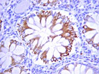
Immunohistochemistry (Paraffin)
Immunocytochemistry
Application Notes
Immunoprecipitation: A previous lot of this antibody was used to detect native and denatured EGFR.
ELISA: A previous lot of this antibody was used in ELISA.
Immunocytochemistry: A previous lot of this antibody was used in IC. Optimal working dilutions must be determined by end user.
Immunohistochemistry(paraffin): Representative testing from a previous lot.
Please refer to the Certificate of Analysis for the lot-specific concentration.
Host
Mouse
Specificity
Reacts strongly and specifically with the intracellular domain of the activated and phosphorylated EGFR in human cell lines and does not react with other phosphorylated proteins.
Isotype
IgG1
Species Reactivity
Human
Mouse
Rat
Species Reactivity Note
Human. Expected to react with mouse and rat based on sequence homology.
The protein encoded by this gene is a transmembrane glycoprotein that is a member of the protein kinase superfamily. This protein is a receptor for members of the epidermal growth factor family. EGFR is a cell surface protein that binds to epidermal growth factor. Binding of the protein to a ligand induces receptor dimerization and tyrosine autophosphorylation and leads to cell proliferation. Mutations in this gene are associated with lung cancer. [provided by RefSeq]
FUNCTION: Receptor for EGF, but also for other members of the EGF family, as TGF-alpha, amphiregulin, betacellulin, heparin-binding EGF-like growth factor, GP30 and vaccinia virus growth factor. Is involved in the control of cell growth and differentiation. Phosphorylates MUC1 in breast cancer cells and increases the interaction of MUC1 with C-SRC and CTNNB1/beta-catenin. FUNCTION: Isoform 2/truncated isoform may act as an antagonist. CATALYTIC ACTIVITY: ATP + a [protein]-L-tyrosine = ADP + a [protein]-L-tyrosine phosphate. SUBUNIT: Binds RIPK1. CBL interacts with the autophosphorylated C-terminal tail of the EGF receptor. Part of a complex with ERBB2 and either PIK3C2A or PIK3C2B. The autophosphorylated form interacts with PIK3C2B, maybe indirectly. Interacts with PELP1. Binds MUC1. SUBCELLULAR LOCATION: Cell membrane; Single-pass type I membrane protein. SUBCELLULAR LOCATION: Isoform 2: Secreted. ALTERNATIVE PRODUCTS: 4 named isoforms [FASTA] produced by alternative splicing.
Molecular Weight
170 kDa
Physicochemical Information
Dimensions
Materials Information
Toxicological Information
Safety Information according to GHS
Safety Information
Product Usage Statements
Quality Assurance
Routinely evaluated by Western Blot on EGF treated A431 lysates. Western Blot Analysis: 1:500 dilution of this lot detected VEGFR on 10 μg of EGF treated A431 lysates.
Usage Statement
Unless otherwise stated in our catalog or other company documentation accompanying the product(s), our products are intended for research use only and are not to be used for any other purpose, which includes but is not limited to, unauthorized commercial uses, in vitro diagnostic uses, ex vivo or in vivo therapeutic uses or any type of consumption or application to humans or animals.
Storage and Shipping Information
Storage Conditions
Stable at -20°C in undiluted aliquots for up to 1 year from date of receipt. Handling Recommendations: Upon first thaw, and prior to removing the cap, centrifuge the vial and gently mix the solution. Aliquot into microcentrifuge tubes and store at -20°C. Avoid repeated freeze/thaw cycles, which may damage IgG and affect product performance. Note: Variability in freezer temperatures below -20°C may cause glycerol containing solutions to become frozen during storage.