Partial restoration of cardiac function with ΔPDZ nNOS in aged mdx model of Duchenne cardiomyopathy.
Lai, Yi, et al.
Hum. Mol. Genet., (2014)
2014
Show Abstract
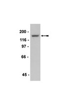
Transgenic gene deletion/over-expression studies have established the cardioprotective role of neuronal nitric oxide synthase (nNOS). However, it remains unclear whether nNOS-mediated heart protection can be translated to gene therapy. In this study, we generated an adeno-associated virus (AAV) nNOS vector and tested its therapeutic efficacy in the aged mdx model of Duchenne cardiomyopathy. A PDZ domain-deleted nNOS gene (ΔPDZ nNOS) was packaged into tyrosine mutant AAV-9 and delivered to the heart of ∼14-month-old female mdx mice, a phenotypic model of Duchenne cardiomyopathy. Seven months later, we observed robust nNOS expression in the myocardium. Supra-physiological ΔPDZ nNOS expression significantly reduced myocardial fibrosis, inflammation and apoptosis. Importantly, electrocardiography and left ventricular hemodynamics were significantly improved in treated mice. Additional studies revealed increased phosphorylation of phospholamban and p70S6K. Collectively, we have demonstrated the therapeutic efficacy of the AAV ΔPDZ nNOS vector in a symptomatic Duchenne cardiomyopathy model. Our results suggest that the cardioprotective role of ΔPDZ nNOS is likely through reduced apoptosis, enhanced phospholamban phosphorylation and improved Akt/mTOR/p70S6K signaling. Our study has opened the door to treat Duchenne cardiomyopathy with ΔPDZ nNOS gene transfer. | | | 24463882
 |
Spinal nNOS regulates phrenic motor facilitation by a 5-HT2B receptor- and NADPH oxidase-dependent mechanism.
MacFarlane, PM; Vinit, S; Mitchell, GS
Neuroscience
269
67-78
2014
Show Abstract
Acute intermittent hypoxia (AIH) induces phrenic long-term facilitation (pLTF) by a mechanism that requires spinal serotonin (5-HT) receptor activation and NADPH oxidase (NOX) activity. Here, we investigated whether: (1) spinal nitric oxide synthase (NOS) activity is necessary for AIH-induced pLTF; (2) episodic exogenous nitric oxide (NO) is sufficient to elicit phrenic motor facilitation (pMF) without AIH (i.e. pharmacologically); and (3) NO-induced pMF requires spinal 5-HT2B receptor and NOX activation. In anesthetized, mechanically ventilated adult male rats, AIH (3 × 5-min episodes; 10% O2; 5 min) elicited a progressive increase in the amplitude of integrated phrenic nerve bursts (i.e. pLTF), which lasted 60 min post-AIH (45.1 ± 8.6% baseline). Pre-treatment with intrathecal (i.t.) injections of a neuronal NOS inhibitor (nNOS-inhibitor-1) near the phrenic motor nucleus attenuated pLTF (14.7 ± 2.5%), whereas an inducible NOS (iNOS) inhibitor (1400 W) had no effect (56.3 ± 8.0%). Episodic i.t. injections (3 × 5μl volume; 5 min) of a NO donor (sodium nitroprusside; SNP) elicited pMF similar in time-course and magnitude (40.4 ± 6.0%, 60 min post-injection) to AIH-induced pLTF. SNP-induced pMF was blocked by a 5-HT2B receptor antagonist (SB206553), a superoxide dismutase mimetic (MnTMPyP), and two NOX inhibitors (apocynin and DPI). Neither pLTF nor pMF was affected by pre-treatment with a protein kinase G (PKG) inhibitor (KT-5823). Thus, spinal nNOS activity is necessary for AIH-induced pLTF, and episodic spinal NO is sufficient to elicit pMF by a mechanism that requires 5-HT2B receptor activation and NOX-derived ROS formation, which indicates AIH (and NO) elicits spinal respiratory plasticity by a nitrergic-serotonergic mechanism. | | | 24680940
|
Effect of octreotide on enteric motor neurons in experimental acute necrotizing pancreatitis.
Zhou, H; Gao, J; Zou, D; Wu, W; Li, Z
PloS one
7
e52163
2012
Show Abstract
Amelioration of intestinal dysmotility and stasis during the early period of acute necrotizing pancreatitis (ANP) appears to be important to reduce the risks of secondary pancreatic infection. We aimed to characterize the association between the neuropathy of the enteric nervous system and gut dysfunction and to examine the effect of octreotide on motor innervation in the early stage of ANP.The rats were randomly divided into eight groups: control+saline; control+octreotide; ANP+saline and ANP+octreotide (24 h, 48 h, 72 h). The spontaneous activity of ileal segments and the response to ACh, l-NNA were recorded. The alterations of myenteric neuronal nitric oxide synthase (nNOS), choline acetyltransferase (CHAT), PGP9.5 and somatostatin receptor 2 (SSTR(2)) immunoreactive cells were evaluated by immunofluorescence and the protein expression of nNOS and CHAT were evaluated by western blot. We found the amplitude of spontaneous contractions at 48 h and the response to ACh at 24 h declined in the ANP+saline rats. A higher contractile response to both ACh and to l-NNA was observed in the ANP+octreotide group, compared with the ANP+saline rats at 24 h. A significant reduction in the nNOS and cholinergic neurons was observed in ANP+saline rats at the three time points. However, this reduction was greatly ameliorated in the presence of octreotide at 24 h and 48 h. The protein expression of CHAT neurons at 24 h and the nNOS neurons at 48 h in the ANP+octreotide rats was much higher than the ANP+saline rats.The pathogenesis of ileus in the early stage of ANP may be related to the neuropathy of the enteric nervous system. Octreotide may reduce the severity of ileus by lessening the damage to enteric motor innervation. | | | 23300603
|
Damage of the interstitial cells of Cajal and myenteric neurons causing ileus in acute necrotizing pancreatitis rats.
Hui Zhou,Li Liu,Yu Bai,Wenbin Wu,Guixiang Li,Jianping Li,Duowu Zou,Jun Gao,Zhaoshen Li
Surgery
149
2011
Show Abstract
Small intestinal motility is impaired in acute necrotizing pancreatitis (ANP). The present study was designed to detect the impairment in small intestinal motility and to assess the role of interstitial cells of Cajal (ICC), myenteric neurons and the associated mechanism in the pathogenesis of ileus during experimentally induced acute pancreatitis. | | | 20570303
|
Lycium barbarum polysaccharides reduce neuronal damage, blood-retinal barrier disruption and oxidative stress in retinal ischemia/reperfusion injury.
Li, SY; Yang, D; Yeung, CM; Yu, WY; Chang, RC; So, KF; Wong, D; Lo, AC
PloS one
6
e16380
2011
Show Abstract
Neuronal cell death, glial cell activation, retinal swelling and oxidative injury are complications in retinal ischemia/reperfusion (I/R) injuries. Lycium barbarum polysaccharides (LBP), extracts from the wolfberries, are good for "eye health" according to Chinese medicine. The aim of our present study is to explore the use of LBP in retinal I/R injury. Retinal I/R injury was induced by surgical occlusion of the internal carotid artery. Prior to induction of ischemia, mice were treated orally with either vehicle (PBS) or LBP (1 mg/kg) once a day for 1 week. Paraffin-embedded retinal sections were prepared. Viable cells were counted; apoptosis was assessed using TUNEL assay. Expression levels of glial fibrillary acidic protein (GFAP), aquaporin-4 (AQP4), poly(ADP-ribose) (PAR) and nitrotyrosine (NT) were investigated by immunohistochemistry. The integrity of blood-retinal barrier (BRB) was examined by IgG extravasations. Apoptosis and decreased viable cell count were found in the ganglion cell layer (GCL) and the inner nuclear layer (INL) of the vehicle-treated I/R retina. Additionally, increased retinal thickness, GFAP activation, AQP4 up-regulation, IgG extravasations and PAR expression levels were observed in the vehicle-treated I/R retina. Many of these changes were diminished or abolished in the LBP-treated I/R retina. Pre-treatment with LBP for 1 week effectively protected the retina from neuronal death, apoptosis, glial cell activation, aquaporin water channel up-regulation, disruption of BRB and oxidative stress. The present study suggests that LBP may have a neuroprotective role to play in ocular diseases for which I/R is a feature. | Immunohistochemistry | Mouse | 21298100
|
Perturbing PSD-95 interactions with NR2B-subtype receptors attenuates spinal nociceptive plasticity and neuropathic pain.
D'Mello, R; Marchand, F; Pezet, S; McMahon, SB; Dickenson, AH
Molecular therapy : the journal of the American Society of Gene Therapy
19
1780-92
2011
Show Abstract
Peripheral inflammation or nerve injury induces a primary afferent barrage into the spinal cord, which can cause N-methyl D-aspartate (NMDA) receptor-dependent alterations in the responses of dorsal horn sensory neurons to subsequent afferent inputs. This plasticity, such as "wind-up" and central sensitization, contributes to the hyperexcitability of dorsal horn neurons and increased pain-related behavior in animal models, as well as clinical signs of chronic pain in humans, hyperalgesia and allodynia. Binding of NMDA receptor subunits by the scaffolding protein postsynaptic density protein-95 (PSD-95) can facilitate downstream intracellular signaling and modulate receptor stability, contributing to synaptic plasticity. Here, we show that spinal delivery of the mimetic peptide Tat-NR2B9c disrupts the interaction between PSD-95 and NR2B subunits in the dorsal horn and selectively reduces NMDA receptor-dependent events including wind-up of spinal sensory neurons, and both persistent formalin-induced neuronal activity and pain-related behaviors, attributed to central sensitization. Furthermore, a single intrathecal injection of Tat-NR2B9c in rats with established nerve injury-induced pain attenuates behavioral signs of mechanical and cold hypersensitivity, with no effect on locomotor performance. Thus, uncoupling of PSD-95 from spinal NR2B-containing NMDA receptors may prevent the neuronal plasticity involved in chronic pain and may be a successful analgesic therapy, reducing side effects associated with receptor blockade. | | | 21427709
|
A study of the spatial protein organization of the postsynaptic density isolated from porcine cerebral cortex and cerebellum.
Yun-Hong, Y; Chih-Fan, C; Chia-Wei, C; Yen-Chung, C
Molecular & cellular proteomics : MCP
10
M110.007138
2011
Show Abstract
Postsynaptic density (PSD) is a protein supramolecule lying underneath the postsynaptic membrane of excitatory synapses and has been implicated to play important roles in synaptic structure and function in mammalian central nervous system. Here, PSDs were isolated from two distinct regions of porcine brain, cerebral cortex and cerebellum. SDS-PAGE and Western blotting analyses indicated that cerebral and cerebellar PSDs consisted of a similar set of proteins with noticeable differences in the abundance of various proteins between these samples. Subsequently, protein localization in these PSDs was analyzed by using the Nano-Depth-Tagging method. This method involved the use of three synthetic reagents, as agarose beads whose surface was covalently linked with a fluorescent, photoactivable, and cleavable chemical crosslinker by spacers of varied lengths. After its application was verified by using a synthetic complex consisting of four layers of different proteins, the Nano-Depth-Tagging method was used here to yield information concerning the depth distribution of various proteins in the PSD. The results indicated that in both cerebral and cerebellar PSDs, glutamate receptors, actin, and actin binding proteins resided in the peripheral regions within ∼ 10 nm deep from the surface and that scaffold proteins, tubulin subunits, microtubule-binding proteins, and membrane cytoskeleton proteins found in mammalian erythrocytes resided in the interiors deeper than 10 nm from the surface in the PSD. Finally, by using the immunoabsorption method, binding partner proteins of two proteins residing in the interiors, PSD-95 and α-tubulin, and those of two proteins residing in the peripheral regions, elongation factor-1α and calcium, calmodulin-dependent protein kinase II α subunit, of cerebral and cerebellar PSDs were identified. Overall, the results indicate a striking similarity in protein organization between the PSDs isolated from porcine cerebral cortex and cerebellum. A model of the molecular structure of the PSD has also been proposed here. | | | 21715321
|
A rat model of smoke inhalation injury: influence of combustion smoke on gene expression in the brain.
Heung M Lee,George H Greeley,David N Herndon,Mala Sinha,Bruce A Luxon,Ella W Englander
Toxicology and applied pharmacology
208
2005
Show Abstract
Acute smoke inhalation causes death and injury in victims of home and industrial fires as well as victims of combat situations. The lethal factors in combustion smoke inhalation are toxic gases and oxygen deficiency, with carbon monoxide (CO) as a primary cause of death. In survivors, inhalation of smoke can result in severe immediate and delayed neuropathologies. To gain insight into the progression of molecular events contributing to smoke inhalation sequelae in the brain, we developed a smoke inhalation rat model and conducted a genome-wide analysis of gene expression. Microarray analysis revealed a modified brain transcriptome with changes peaking at 24 h and subsiding within 7 days post-smoke. Overall, smoke inhalation down regulated genes associated with synaptic function, neurotransmission, and neurotrophic support, and upregulated genes associated with stress responses, including nitric oxide synthesis, antioxidant defenses, proteolysis, inflammatory response, and glial activation. Notably, among the affected genes, many have been previously implicated in other types of brain injury, demonstrating the usefulness of microarrays for analysis of changes in gene expression in complex insults. In accord with previously described modulations of nitric oxide homeostasis in CO poisoning, microarray analysis revealed increased brain expression of nitric oxide synthase (NOS) and NOS ligand after inhalation of smoke. Furthermore, immunostaining showed significant elevations in perivascular NOS and in protein nitration, corroborating the involvement of nitric oxide perturbations in post-smoke sequelae in the brain. Thus, the new rat model, in combination with microarray analyses, affords insight into the complex molecular pathophysiology of smoke inhalation in the brain. | | | 15893353
|
The DDAH/ADMA/NOS pathway.
Tran, Cam T L, et al.
Atherosclerosis. Supplements, 4: 33-40 (2003)
2003
Show Abstract
An increasing number of reports in the literature indicate that endogenously produced inhibitors of nitric oxide synthase (NOS), particularly asymmetric dimethylarginine (ADMA) regulate nitric oxide generation in numerous disease states. Two dimethylarginine dimethylaminohydrolase (DDAH) enzymes metabolise ADMA. We and others have postulated that activity of DDAH is a key determinant of ADMA levels in vivo. This review summarises recent advances in the regulation and function of DDAH enzymes and its role in the regulation of nitric oxide generation. | | | 14664901
|
AMPK signaling in contracting human skeletal muscle: acetyl-CoA carboxylase and NO synthase phosphorylation.
Chen, Z P, et al.
Am. J. Physiol. Endocrinol. Metab., 279: E1202-6 (2000)
2000
Show Abstract
AMP-activated protein kinase (AMPK) is a metabolic stress-sensing protein kinase responsible for coordinating metabolism and energy demand. In rodents, exercise accelerates fatty acid metabolism, enhances glucose uptake, and stimulates nitric oxide (NO) production in skeletal muscle. AMPK phosphorylates and inhibits acetyl-coenzyme A (CoA) carboxylase (ACC) and enhances GLUT-4 translocation. It has been reported that human skeletal muscle malonyl-CoA levels do not change in response to exercise, suggesting that other mechanisms besides inhibition of ACC may be operating to accelerate fatty acid oxidation. Here, we show that a 30-s bicycle sprint exercise increases the activity of the human skeletal muscle AMPK-alpha1 and -alpha2 isoforms approximately two- to threefold and the phosphorylation of ACC at Ser(79) (AMPK phosphorylation site) approximately 8.5-fold. Under these conditions, there is also an approximately 5.5-fold increase in phosphorylation of neuronal NO synthase-mu (nNOSmu;) at Ser(1451). These observations support the concept that inhibition of ACC is an important component in stimulating fatty acid oxidation in response to exercise and that there is coordinated regulation of nNOSmu to protect the muscle from ischemia/metabolic stress. | | | 11052978
|