Si cierra, no se guardará su personalización salvo que haya añadido el artículo a su carrito de la compra o a favoritos.
Pulse OK para cerrar la herramienta MILLIPLEX® MAP o Cancelar para volver a su selección.
Elija paneles personalizables y kits premezclos - O - MAPmates™ de señalización celular
Diseñe y calcule el precio de sus kits MILLIPLEX® MAP.
Paneles personalizados y kits premezclados
Nuestra amplia cartera de productos consta de paneles multiplex que le permiten elegir, dentro del panel, los analitos que mejor se ajustan a sus requisitos. En una pestaña distinta puede elegir el formato de citocina premezclada o un kit single plex.
Kits de señalización celular y MAPmates™
Elija los kits preparados para poder explorar las vías o los procesos enteros. O diseñe sus propios kits eligiendo single plex MAPmates™ según las directrices proporcionadas.
No deben combinarse los siguientes MAPmates™: -MAPmates™ que requieren un tampón de ensayo diferente. -Pares MAPmate™ fosfoespecíficos y totales, por ejemplo, GSK3β y GSK3β (Ser 9). -MAPmates™ con panTyr y específicos de sitio; por ejemplo, receptor del fosfo-EGF y fosfo-STAT1 (Tyr701). -Más de 1 fosfo-MAPmate™ para una sola diana (Akt, STAT3). -La GAPDH y la β-tubulina no pueden combinarse con kits o MAPmates™ que contengan panTyr.
.
Número de referencia
Descripción para pedidos
Cant./Env.
Lista
Este artículo se ha añadido a favoritos.
Seleccione una especie, un tipo de panel, un kit o un tipo de muestra
Para empezar a diseñar su kit MILLIPLEX® MAP, seleccione una especie, un tipo de panel o un kit de interés.
Custom Premix Selecting "Custom Premix" option means that all of the beads you have chosen will be premixed in manufacturing before the kit is sent to you.
Catalogue Number
Ordering Description
Qty/Pack
List
Este artículo se ha añadido a favoritos.
Especie
Tipo de panel
Kit seleccionado
Cant.
Número de referencia
Descripción para pedidos
Cant./Env.
Precio de catálogo
96-Well Plate
Cant.
Número de referencia
Descripción para pedidos
Cant./Env.
Precio de catálogo
Añadir más reactivos (Se necesita tampón y un kit de detección para usar con MAPmates)
Cant.
Número de referencia
Descripción para pedidos
Cant./Env.
Precio de catálogo
48-602MAG
Buffer Detection Kit for Magnetic Beads
1 Kit
Opción para ahorrar espacio Los clientes que adquieran múltiples kits pueden optar por ahorrar espacio de almacenamiento retirando el embalaje del kit y recibiendo los componentes de sus ensayos multiplex en bolsas de plástico para un almacenamiento más compacto.
Este artículo se ha añadido a favoritos.
El producto se ha añadido a su carrito
Ahora puede personalizar otro kit, elegir un kit premezclado, tramitarlo o cerrar la herramienta de pedidos.
Detect EGFR (cytoplasmic domain) using this Anti-EGFR (cytoplasmic domain) Antibody, clone 8G6.2 validated for use in IC, IH, IP & WB.
More>>Detect EGFR (cytoplasmic domain) using this Anti-EGFR (cytoplasmic domain) Antibody, clone 8G6.2 validated for use in IC, IH, IP & WB. Less<<
Anti-EGFR (cytoplasmic domain) Antibody, clone 8G6.2: Ficha de datos de seguridad (MSDS o SDS), certificado de análisis y de calidad (CoA y CoQ), expedientes, folletos y otros documentos disponibles.
The epidermal growth factor receptor (EGFR; ErbB-1; HER1 in humans) is the cell-surface receptor for members of the epidermal growth factor family (EGF-family) of extracellular protein ligands. The epidermal growth factor receptor is a member of the ErbB family of receptors, a subfamily of four closely related receptor tyrosine kinases: EGFR (ErbB-1), HER2/c-neu (ErbB-2), Her 3 (ErbB-3) and Her 4 (ErbB-4). Mutations affecting EGFR expression or activity could result in cancer. EGFR (epidermal growth factor receptor) exists on the cell surface and is activated by binding of its specific ligands, including epidermal growth factor and transforming growth factor alpha (TGFα). Upon activation by its growth factor ligands, EGFR undergoes a transition from an inactive monomeric form to an active homodimer. In addition to forming homodimers after ligand binding, EGFR may pair with another member of the ErbB receptor family, such as ErbB2/Her2/neu, to create an activated heterodimer. There is also evidence to suggest that clusters of activated EGFRs form, although it remains unclear whether this clustering is important for activation itself or occurs subsequent to activation of individual dimers. EGFR dimerization stimulates its intrinsic intracellular protein-tyrosine kinase activity. As a result, autophosphorylation of five tyrosine (Y) residues in the C-terminal domain of EGFR occurs. These are Y992, Y1045, Y1068, Y1148 and Y1173. This autophosphorylation elicits downstream activation and signaling by several other proteins that associate with the phosphorylated tyrosines through their own phosphotyrosine-binding SH2 domains. These downstream signaling proteins initiate several signal transduction cascades, principally the MAPK, Akt and JNK pathways, leading to DNA synthesis and cell proliferation. Such proteins modulate phenotypes such as cell migration, adhesion, and proliferation. The kinase domain of EGFR can also cross-phosphorylate tyrosine residues of other receptors it is aggregated with, and can itself be activated in that manner.
References
Product Information
Format
Purified
Presentation
Purified mouse monoclonal antibody in buffer containing 0.1 M Tris-Glycine (pH 7.4), 150 mM NaCl with 0.05% sodium azide.
Detect EGFR (cytoplasmic domain) using this Anti-EGFR (cytoplasmic domain) Antibody, clone 8G6.2 validated for use in IC, IH, IP & WB.
Key Applications
Immunocytochemistry
Immunohistochemistry
Immunoprecipitation
Western Blotting
Application Notes
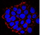
Immunocytochemistry Analysis: A431 cells were grown, fixed, permeablized, and stained with anti-EGFR, clone 8G6.2
Confocal Immunocytochemistry Analysis: A431 cells were grown, fixed, permeablized, and stained with anti-EGFR, clone 8G6.2
Immunoprecipitation: 100 μg of A431 whole cell lysate was lysed in RIPA buffer and immunoprecipitated (IP) with 5 μg of Anti-EGFR, cytoplasmic domain, clone 8G6.2
Immunohistochemistry Analysis: Tissue was stained with anti-EGFR, cytoplasmic domain, clone 8G6.2 at a 1:100 dilution and IHC-Select Detection with HRP-DAB reagents.
Biological Information
Immunogen
Recombinant protein encoding the cytoplasmic domain (C-terminal) of human EGF Receptor.
Epitope
Cytoplasmic Domain
Clone
8G6.2
Concentration
Please refer to the Certificate of Analysis for the lot-specific concentration.
Host
Mouse
Specificity
This antibody detects the cytoplasmic domain of EGFR.
Isotype
IgG1κ
Species Reactivity
Human
Mouse
Rat
Species Reactivity Note
Evaluated by western blot on A431 cell lysate Western Blot Analysis: 1:2,000 dilution of this antibody was used to detect EGFR in A431 cell lysate.
The protein encoded by this gene is a transmembrane glycoprotein that is a member of the protein kinase superfamily. This protein is a receptor for members of the epidermal growth factor family. EGFR is a cell surface protein that binds to epidermal growth factor. Binding of the protein to a ligand induces receptor dimerization and tyrosine autophosphorylation and leads to cell proliferation. Mutations in this gene are associated with lung cancer. [provided by RefSeq].
FUNCTION: SwissProt: P00533 # Isoform 2/truncated isoform may act as an antagonist. SIZE: 1210 amino acids; 134277 Da SUBUNIT: Binds RIPK1. CBL interacts with the autophosphorylated C- terminal tail of the EGF receptor. Part of a complex with ERBB2 and either PIK3C2A or PIK3C2B. The autophosphorylated form interacts with PIK3C2B, maybe indirectly. Interacts with PELP1. SUBCELLULAR LOCATION: Cell membrane; Single-pass type I membrane protein. & Isoform 2: Secreted. TISSUE SPECIFICITY: Expressed in placenta. Isoform 2 is also expressed in ovarian cancers. PTM: Phosphorylation of Ser-695 is partial and occurs only if Thr- 693 is phosphorylated. & Monoubiquitinated and polyubiquitinated upon EGF stimulation; which does not affect tyrosine kinase activity or signaling capacity but may play a role in lysosomal targeting. Polyubiquitin linkage is mainly through 'Lys-63', but linkage through 'Lys-48', 'Lys-11' and 'Lys-29' also occur.DISEASE:SwissProt: P00533 # Defects in EGFR are associated with lung cancer [MIM:211980]. SIMILARITY: SwissProt: P00533 ## Belongs to the protein kinase superfamily. Tyr protein kinase family. EGF receptor subfamily. & Contains 1 protein kinase domain. MISCELLANEOUS: Binding of EGF to the receptor leads to dimerization, internalization of the EGF-receptor complex, induction of the tyrosine kinase activity, stimulation of cell DNA synthesis, and cell proliferation.
Molecular Weight
180 kDa
Physicochemical Information
Dimensions
Materials Information
Toxicological Information
Safety Information according to GHS
Safety Information
Product Usage Statements
Quality Assurance
Evaluated by western blot on A431 cell lysate Western Blot Analysis: 1:2,000 dilution of this antibody was used to detect EGFR in A431 cell lysate.
Usage Statement
Unless otherwise stated in our catalog or other company documentation accompanying the product(s), our products are intended for research use only and are not to be used for any other purpose, which includes but is not limited to, unauthorized commercial uses, in vitro diagnostic uses, ex vivo or in vivo therapeutic uses or any type of consumption or application to humans or animals.