Potential role for kv3.1b channels as oxygen sensors.
Osipenko, O N, et al.
Circ. Res., 86: 534-40 (2000)
2000
Show Abstract
Hypoxia inhibits voltage-gated K channels in pulmonary artery smooth muscle (PASM). This is thought to contribute to hypoxic pulmonary vasoconstriction by promoting membrane depolarization, Ca(2+) influx, and contraction. Several of the K-channel subtypes identified in pulmonary artery have been implicated in the response to hypoxia, but contradictory evidence clouds the identity of the oxygen-sensing channels. Using patch-clamp techniques, this study investigated the effect of hypoxia on recombinant Kv1 channels previously identified in pulmonary artery (Kv1.1, Kv1.2, and Kv1.5) and Kv3.1b, which has similar kinetic and pharmacological properties to native oxygen-sensitive currents. Hypoxia failed to inhibit any Kv1 channel, but it inhibited Kv3.1b channels expressed in L929 cells, as shown by a reduction of whole-cell current and single-channel activity, without affecting unitary conductance. Inhibition was retained in excised membrane patches, suggesting a membrane-delimited mechanism. Using reverse transcription-polymerase chain reaction and immunocytochemistry, Kv3.1b expression was demonstrated in PASM cells. Moreover, hypoxia inhibited a K(+) current in rabbit PASM cells in the presence of charybdotoxin and capsaicin, which preserve Kv3.1b while blocking most other Kv channels, but not in the presence of millimolar tetraethylammonium ions, which abolish Kv3.1b current. Kv3.1b channels may therefore contribute to oxygen sensing in pulmonary artery. | 10720415
 |
Angiotensin II type 1 receptor-mediated inhibition of K+ channel subunit kv2.2 in brain stem and hypothalamic neurons.
Gelband, C H, et al.
Circ. Res., 84: 352-9 (1999)
1999
Show Abstract
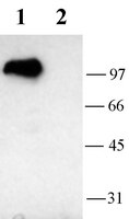
Angiotensin II (Ang II) has powerful modulatory actions on cardiovascular function that are mediated by specific receptors located on neurons within the hypothalamus and brain stem. Incubation of neuronal cocultures of rat hypothalamus and brain stem with Ang II elicits an Ang II type 1 (AT1) receptor-mediated inhibition of total outward K+ current that contributes to an increase in neuronal firing rate. However, the exact K+ conductance(s) that is inhibited by Ang II are not established. Pharmacological manipulation of total neuronal outward K+ current revealed a component of K+ current sensitive to quinine, tetraethylammonium, and 4-aminopyridine, with IC50 values of 21.7 micromol/L, 1.49 mmol/L, and 890 micromol/L, respectively, and insensitive to alpha-dendrotoxin (100 to 500 nmol/L), charybdotoxin (100 to 500 nmol/L), and mast cell degranulating peptide (1 micromol/L). Collectively, these data suggest the presence of Kv2.2 and Kv3.1b. Biophysical examination of the quinine-sensitive neuronal K+ current demonstrated a macroscopic conductance with similar biophysical properties to those of Kv2.2 and Kv3.1b. Ang II (100 nmol/L), in the presence of the AT2 receptor blocker PD123,319, elicited an inhibition of neuronal K+ current that was abolished by quinine (50 micromol/L). Reverse transcriptase-polymerase chain reaction analysis confirmed the presence of Kv2.2 and Kv3.1b mRNA in these neurons. However, Western blot analyses demonstrated that only Kv2.2 protein was present. Coexpression of Kv2.2 and the AT1 receptor in Xenopus oocytes demonstrated an Ang II-induced inhibition of Kv2.2 current. Therefore, these data suggest that inhibition of Kv2.2 contributes to the AT1 receptor-mediated reduction of neuronal K+ current and subsequently to the modulation of cardiovascular function. | 10024310
|
Cortical neurons immunoreactive for the potassium channel Kv3.1b subunit are predominantly surrounded by perineuronal nets presumed as a buffering system for cations.
Härtig, W, et al.
Brain Res., 842: 15-29 (1999)
1999
Show Abstract
Perineuronal nets (PNs) are known as chondroitin sulphate-rich, lattice-like coatings of the extracellular matrix. In the cortex of mammalian species investigated so far, they were mainly found around GABAergic neurons, but to a lesser degree also around pyramidal cells. Previous investigations in the rat revealed similar distribution patterns of fast-firing neurons expressing both the Kv3.1b subunit of voltage-gated potassium channels and the calcium-binding protein parvalbumin. In the present study, triple fluorescence labelling was applied for the simultaneous demonstration of PNs with the N-acetylgalactosamine-specific Wisteria floribunda agglutinin (WFA), parvalbumin-immunoreactivity (ir) with a monoclonal antibody and of Kv3.1b-ir with several rabbit antibodies. Subsets of non-pyramidal neurons - enwrapped by PNs and expressing parvalbumin and Kv3.1b - were detected in the rat and monkey neocortex and hippocampus. In the rat, faintly stained PNs were additionally found around several layer II/III and V pyramidal cells immunonegative for Kv3.1b, but contacted by Kv3.1b-containing boutons. In the monkey, more intensely labelled PNs frequently occurred around pyramidal cells which themselves appeared to be Kv3. 1b-immunopositive. We also observed minor Kv3.1b-ir and parvalbumin-ir cortical cell populations which were devoid of PNs; occasionally, nets were detected around neurons lacking both immunoreactivities. By confocal laser scanning microscopy, Kv3.1b-ir and WFA-binding sites were found adjoining at the soma and proximal dendritic surface, while lectin-binding sites usually extended on more distal dendritic segments and the axon initial segments which failed to express detectable Kv3.1b-ir. This spatial relationship of both markers was also confirmed by combined WFA-gold labelling and Kv3.1b-immunoperoxidase staining at the electron microscopic level. The data are used for a critical examination of current hypotheses concerning the functional role of PNs. We conclude that PNs may serve as rapid local buffers of excess cation changes in the extracellular space. Somatic membranes of fast-spiking neurons seem to be a main, but not the only source of such changes. | 10526091
|
Heteromultimeric delayed-rectifier K+ channels in schwann cells: developmental expression and role in cell proliferation.
Sobko, A, et al.
J. Neurosci., 18: 10398-408 (1998)
1998
Show Abstract
Schwann cells (SCs) are responsible for myelination of nerve fibers in the peripheral nervous system. Voltage-dependent K+ currents, including inactivating A-type (KA), delayed-rectifier (KD), and inward-rectifier (KIR) K+ channels, constitute the main conductances found in SCs. Physiological studies have shown that KD channels may play an important role in SC proliferation and that they are downregulated in the soma as proliferation ceases and myelination proceeds. Recent studies have begun to address the molecular identity of K+ channels in SCs. Here, we show that a large repertoire of K+ channel alpha subunits of the Shaker (Kv1.1, Kv1.2, Kv1.4, and Kv1.5), Shab (Kv2.1), and Shaw (Kv3.1b and Kv3.2) families is expressed in mouse SCs and sciatic nerve. We characterized heteromultimeric channel complexes that consist of either Kv1.5 and Kv1.2 or Kv1.5 and Kv1.4. In postnatal day 4 (P4) sciatic nerve, most of the Kv1.2 channel subunits are involved in heteromultimeric association with Kv1.5. Despite the presence of Kv1. 1 and Kv1.2 alpha subunits, the K+ currents were unaffected by dendrotoxin I (DTX), suggesting that DTX-sensitive channel complexes do not account substantially for SC KD currents. SC proliferation was found to be potently blocked by quinidine or 4-aminopyridine but not by DTX. Consistent with previous physiological studies, our data show that there is a marked downregulation of all KD channel alpha subunits from P1-P4 to P40 in the sciatic nerve. Our results suggest that KD currents are accounted for by a complex combinatorial activity of distinct K+ channel complexes and confirm that KD channels are involved in SC proliferation. | 9852577
|