Nicotine accelerates diabetes-induced retinal changes.
Boretsky, A; Gupta, P; Tirgan, N; Liu, R; Godley, BF; Zhang, W; Tilton, RG; Motamedi, M
Current eye research
40
368-77
2015
Show Abstract
To investigate the effects of nicotine on retinal alterations in early-stage diabetes in an established rodent model.Sprague-Dawley rats were examined using a combination of confocal scanning laser ophthalmoscopy and spectral domain optical coherence tomography to determine changes in retinal structure in response to nicotine exposure, diabetes and the combined effects of nicotine and diabetes. Diabetes was induced by a single injection of 65 mg/kg streptozotocin and nicotine injections were administered subcutaneously daily. Retinal thickness in the superior, inferior, nasal and temporal quadrants were determined based on the spectral domain optical coherence tomography (SD-OCT) volume scans (20° × 20°) centered on the optic disc. Segmentation of discrete retinal layers was performed on a subset of SD-OCT cross-sections to further examine changes in each treatment group. Survival of neurons within the ganglion cell layer (GCL) was assessed by confocal morphometric imaging.The control group did not experience any significant change throughout the study. The nicotine treatment group experienced an average decrease in total retinal thickness (TRT) of 9.4 µm with the majority of the loss localized within the outer nuclear layer (ONL) as determined by segmentation analysis (p less than 0.05). The diabetic group exhibited a trend toward decreased TRT while segmentation analysis of the diabetic retinopathy (DR) group revealed significant thinning within the ONL (p less than 0.05). The combination of nicotine and diabetes revealed a significant increase of 8.9 µm in the TRT (p less than 0.05) accompanied by a decrease in the number of GCL neurons.We demonstrated significant temporal changes in retinal morphology in response to nicotine exposure, diabetes and with the combined effects of nicotine and diabetes. These findings may have implications in determining treatment strategies for diabetic patients using products containing nicotine, such as cigarettes, smokeless tobacco, electronic cigarettes or smoking cessation products. | | | 24911405
 |
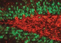
An anterograde rabies virus vector for high-resolution large-scale reconstruction of 3D neuron morphology.
Haberl, MG; Viana da Silva, S; Guest, JM; Ginger, M; Ghanem, A; Mulle, C; Oberlaender, M; Conzelmann, KK; Frick, A
Brain structure & function
220
1369-79
2015
Show Abstract
Glycoprotein-deleted rabies virus (RABV ∆G) is a powerful tool for the analysis of neural circuits. Here, we demonstrate the utility of an anterograde RABV ∆G variant for novel neuroanatomical approaches involving either bulk or sparse neuronal populations. This technology exploits the unique features of RABV ∆G vectors, namely autonomous, rapid high-level expression of transgenes, and limited cytotoxicity. Our vector permits the unambiguous long-range and fine-scale tracing of the entire axonal arbor of individual neurons throughout the brain. Notably, this level of labeling can be achieved following infection with a single viral particle. The vector is effective over a range of ages (greater than 14 months) aiding the studies of neurodegenerative disorders or aging, and infects numerous cell types in all brain regions tested. Lastly, it can also be readily combined with retrograde RABV ∆G variants. Together with other modern technologies, this tool provides new possibilities for the investigation of the anatomy and physiology of neural circuits. | | | 24723034
|
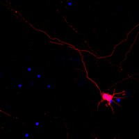
Pre-existing astrocytes form functional perisynaptic processes on neurons generated in the adult hippocampus.
Krzisch, M; Temprana, SG; Mongiat, LA; Armida, J; Schmutz, V; Virtanen, MA; Kocher-Braissant, J; Kraftsik, R; Vutskits, L; Conzelmann, KK; Bergami, M; Gage, FH; Schinder, AF; Toni, N
Brain structure & function
220
2027-42
2015
Show Abstract
The adult dentate gyrus produces new neurons that morphologically and functionally integrate into the hippocampal network. In the adult brain, most excitatory synapses are ensheathed by astrocytic perisynaptic processes that regulate synaptic structure and function. However, these processes are formed during embryonic or early postnatal development and it is unknown whether astrocytes can also ensheathe synapses of neurons born during adulthood and, if so, whether they play a role in their synaptic transmission. Here, we used a combination of serial-section immuno-electron microscopy, confocal microscopy, and electrophysiology to examine the formation of perisynaptic processes on adult-born neurons. We found that the afferent and efferent synapses of newborn neurons are ensheathed by astrocytic processes, irrespective of the age of the neurons or the size of their synapses. The quantification of gliogenesis and the distribution of astrocytic processes on synapses formed by adult-born neurons suggest that the majority of these processes are recruited from pre-existing astrocytes. Furthermore, the inhibition of astrocytic glutamate re-uptake significantly reduced postsynaptic currents and increased paired-pulse facilitation in adult-born neurons, suggesting that perisynaptic processes modulate synaptic transmission on these cells. Finally, some processes were found intercalated between newly formed dendritic spines and potential presynaptic partners, suggesting that they may also play a structural role in the connectivity of new spines. Together, these results indicate that pre-existing astrocytes remodel their processes to ensheathe synapses of adult-born neurons and participate to the functional and structural integration of these cells into the hippocampal network. | | | 24748560
|
Quantitative and functional interrogation of parent-of-origin allelic expression biases in the brain.
Perez, JD; Rubinstein, ND; Fernandez, DE; Santoro, SW; Needleman, LA; Ho-Shing, O; Choi, JJ; Zirlinger, M; Chen, SK; Liu, JS; Dulac, C
eLife
4
e07860
2015
Show Abstract
The maternal and paternal genomes play different roles in mammalian brains as a result of genomic imprinting, an epigenetic regulation leading to differential expression of the parental alleles of some genes. Here we investigate genomic imprinting in the cerebellum using a newly developed Bayesian statistical model that provides unprecedented transcript-level resolution. We uncover 160 imprinted transcripts, including 41 novel and independently validated imprinted genes. Strikingly, many genes exhibit parentally biased--rather than monoallelic--expression, with different magnitudes according to age, organ, and brain region. Developmental changes in parental bias and overall gene expression are strongly correlated, suggesting combined roles in regulating gene dosage. Finally, brain-specific deletion of the paternal, but not maternal, allele of the paternally-biased Bcl-x, (Bcl2l1) results in loss of specific neuron types, supporting the functional significance of parental biases. These findings reveal the remarkable complexity of genomic imprinting, with important implications for understanding the normal and diseased brain. | | | 26140685
|
Brain Localization and Neurotoxicity Evaluation of Polysorbate 80-Modified Chitosan Nanoparticles in Rats.
Yuan, ZY; Hu, YL; Gao, JQ
PloS one
10
e0134722
2015
Show Abstract
The toxicity evaluation of inorganic nanoparticles has been reported by an increasing number of studies, but toxicity studies concerned with biodegradable nanoparticles, especially the neurotoxicity evaluation, are still limited. For example, the potential neurotoxicity of Polysorbate 80-modified chitosan nanoparticles (Tween 80-modified chitosan nanoparticles, TmCS-NPs), one of the most widely used brain targeting vehicles, remains unknown. In the present study, TmCS-NPs with a particle size of 240 nm were firstly prepared by ionic cross-linking of chitosan with tripolyphosphate. Then, these TmCS-NPs were demonstrated to be entered into the brain and specially deposited in the frontal cortex and cerebellum after systemic injection. Moreover, the concentration of TmCS-NPs in these two regions was found to decrease over time. Although no obvious changes were observed for oxidative stress in the in vivo rat model, the body weight was found to remarkably decreased in a dose-dependent manner after exposure to TmCS-NPs for seven days. Besides, apoptosis and necrosis of neurons, slight inflammatory response in the frontal cortex, and decrease of GFAP expression in the cerebellum were also detected in mouse injected with TmCS-NPs. This study is the first report on the sub-brain biodistribution and neurotoxicity studies of TmCS-NPs. Our results provide new insights into the toxicity evaluation of nanoparticles and our findings would help contribute to a better understanding of the neurotoxicity of biodegradable nanomaterials used in pharmaceutics. | | | 26248340
|
Taenia solium: Development of an Experimental Model of Porcine Neurocysticercosis.
Fleury, A; Trejo, A; Cisneros, H; García-Navarrete, R; Villalobos, N; Hernández, M; Villeda Hernández, J; Hernández, B; Rosas, G; Bobes, RJ; de Aluja, AS; Sciutto, E; Fragoso, G
PLoS neglected tropical diseases
9
e0003980
2015
Show Abstract
Human neurocysticercosis (NC) is caused by the establishment of Taenia solium larvae in the central nervous system. NC is a severe disease still affecting the population in developing countries of Latin America, Asia, and Africa. While great improvements have been made on NC diagnosis, treatment, and prevention, the management of patients affected by extraparenchymal parasites remains a challenge. The development of a T. solium NC experimental model in pigs that will allow the evaluation of new therapeutic alternatives is herein presented. Activated oncospheres (either 500 or 1000) were surgically implanted in the cerebral subarachnoid space of piglets. The clinical status and the level of serum antibodies in the animals were evaluated for a 4-month period after implantation. The animals were sacrificed, cysticerci were counted during necropsy, and both the macroscopic and microscopic characteristics of cysts were described. Based on the number of established cysticerci, infection efficiency ranged from 3.6% (1000 oncospheres) to 5.4% (500 oncospheres). Most parasites were caseous or calcified (38/63, 60.3%) and were surrounded by an exacerbated inflammatory response with lymphocyte infiltration and increased inflammatory markers. The infection elicited specific antibodies but no neurological signs. This novel experimental model of NC provides a useful tool to evaluate new cysticidal and anti-inflammatory approaches and it should improve the management of severe NC patients, refractory to the current treatments. | | | 26252878
|
TWIK-Related Spinal Cord K⁺ Channel Expression Is Increased in the Spinal Dorsal Horn after Spinal Nerve Ligation.
Hwang, HY; Zhang, E; Park, S; Chung, W; Lee, S; Kim, DW; Ko, Y; Lee, W
Yonsei medical journal
56
1307-15
2015
Show Abstract
The TWIK-related spinal cord K⁺ channel (TRESK) has recently been discovered and plays an important role in nociceptor excitability in the pain pathway. Because there have been no reports on the TRESK expression or its function in the dorsal horn of the spinal cord in neuropathic pain, we analyzed TRESK expression in the spinal dorsal horn in a spinal nerve ligation (SNL) model.We established a SNL mouse model by using the L5-6 spinal nerves ligation. We used real-time polymerase chain reaction and immunohistochemistry to investigate TRESK expression in the dorsal horn and L5 dorsal rot ganglion (DRG).The SNL group showed significantly higher expression of TRESK in the ipsilateral dorsal horn under pain, but low expression in L5 DRG. Double immunofluorescence staining revealed that immunoreactivity of TRESK was mostly restricted in neuronal cells, and that synapse markers GAD67 and VGlut2 appeared to be associated with TRESK expression. We were unable to find a significant association between TRESK and calcineurin by double immunofluorescence.TRESK in spinal cord neurons may contribute to the development of neuropathic pain following injury. | | | 26256973
|
Vibrissa Self-Motion and Touch Are Reliably Encoded along the Same Somatosensory Pathway from Brainstem through Thalamus.
Moore, JD; Mercer Lindsay, N; Deschênes, M; Kleinfeld, D
PLoS biology
13
e1002253
2015
Show Abstract
Active sensing involves the fusion of internally generated motor events with external sensation. For rodents, active somatosensation includes scanning the immediate environment with the mystacial vibrissae. In doing so, the vibrissae may touch an object at any angle in the whisk cycle. The representation of touch and vibrissa self-motion may in principle be encoded along separate pathways, or share a single pathway, from the periphery to cortex. Past studies established that the spike rates in neurons along the lemniscal pathway from receptors to cortex, which includes the principal trigeminal and ventral-posterior-medial thalamic nuclei, are substantially modulated by touch. In contrast, spike rates along the paralemniscal pathway, which includes the rostral spinal trigeminal interpolaris, posteromedial thalamic, and ventral zona incerta nuclei, are only weakly modulated by touch. Here we find that neurons along the lemniscal pathway robustly encode rhythmic whisking on a cycle-by-cycle basis, while encoding along the paralemniscal pathway is relatively poor. Thus, the representations of both touch and self-motion share one pathway. In fact, some individual neurons carry both signals, so that upstream neurons with a supralinear gain function could, in principle, demodulate these signals to recover the known decoding of touch as a function of vibrissa position in the whisk cycle. | | | 26393890
|
Forebrain microglia from wild-type but not adult 5xFAD mice prevent amyloid-β plaque formation in organotypic hippocampal slice cultures.
Hellwig, S; Masuch, A; Nestel, S; Katzmarski, N; Meyer-Luehmann, M; Biber, K
Scientific reports
5
14624
2015
Show Abstract
The role of microglia in amyloid-β (Aβ) deposition is controversial. In the present study, an organotypic hippocampal slice culture (OHSC) system with an in vivo-like microglial-neuronal environment was used to investigate the potential contribution of microglia to Aβ plaque formation. We found that microglia ingested Aβ, thereby preventing plaque formation in OHSCs. Conversely, Aβ deposits formed rapidly in microglia-free wild-type slices. The capacity to prevent Aβ plaque formation was absent in forebrain microglia from young adult but not juvenile 5xFamilial Alzheimer's disease (FAD) mice. Since no loss of Aβ clearance capacity was observed in both wild-type and cerebellar microglia from 5xFAD animals, the high Aβ1-42 burden in the forebrain of 5xFAD animals likely underlies the exhaustion of microglial Aβ clearance capacity. These data may therefore explain why Aβ plaque formation has never been described in wild-type mice, and point to a beneficial role of microglia in AD pathology. We also describe a new method to study Aβ plaque formation in a cell culture setting. | | | 26416689
|
Hepatic but Not CNS-Expressed Human C-Reactive Protein Inhibits Experimental Autoimmune Encephalomyelitis in Transgenic Mice.
Wright, TT; Jimenez, RV; Morgan, TE; Bali, N; Hou, X; McCrory, MA; Finch, CE; Szalai, AJ
Autoimmune diseases
2015
640171
2015
Show Abstract
We recently demonstrated that human C-reactive protein (CRP), expressed hepatically in transgenic mice (CRPtg), improved the outcome of experimental autoimmune encephalomyelitis (EAE), a murine model of multiple sclerosis (MS). The liver is the primary site of CRP synthesis in humans and in CRPtg mice but is also expressed by both at low levels in the CNS. To determine if CNS expression of human CRP is sufficient to impact EAE, we generated neuronal CRP transgenic mice (nCRPtg) wherein human CRP expression is driven by the neuron-specific Ca(2+)/calmodulin-dependent protein kinase IIα (CaMKIIα) gene promoter. We found that hepatically expressed/blood-borne CRP, but not CNS expressed CRP, lessened EAE severity. These outcomes indicate that the protective actions of human CRP in EAE are manifested in the periphery and not in the CNS and reveal a previously unappreciated site specificity for the beneficial actions of CRP in CNS disease. | | | 26421184
|