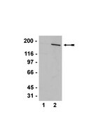
PKB-Mediated Thr649 Phosphorylation of AS160/TBC1D4 Regulates the R-Wave Amplitude in the Heart.
Quan, C; Xie, B; Wang, HY; Chen, S
PloS one
10
e0124491
2015
Mostrar resumen
The Rab GTPase activating protein (RabGAP), AS160/TBC1D4, is an important substrate of protein kinase B (PKB), and regulates insulin-stimulated trafficking of glucose transporter 4. Besides, AS160/TBC1D4 has also been shown to regulate trafficking of many other membrane proteins including FA translocase/CD36 in cardiomyocytes. However, it is not clear whether it plays any role in regulating heart functions in vivo. Here, we found that PKB-mediated phosphorylation of Thr649 on AS160/TBC1D4 represented one of the major PAS-binding signals in the heart in response to insulin. Mutation of Thr649 to a non-phosphorylatable alanine increased the R-wave amplitude in the AS160Thr649Ala knockin mice. However, this knockin mutation did not affect the heart functions under both normal and infarct conditions. Interestingly, myocardial infarction induced the expression of a related RabGAP, TBC1D1, in the infarct zone as well as in the border zone. Together, these data show that the Thr649 phosphorylation of AS160/TBC1D4 plays an important role in the heart's electrical conduction system through regulating the R-wave amplitude. | | | 25923736
 |
Acute mTOR inhibition induces insulin resistance and alters substrate utilization in vivo.
Kleinert, M; Sylow, L; Fazakerley, DJ; Krycer, JR; Thomas, KC; Oxbøll, AJ; Jordy, AB; Jensen, TE; Yang, G; Schjerling, P; Kiens, B; James, DE; Ruegg, MA; Richter, EA
Molecular metabolism
3
630-41
2014
Mostrar resumen
The effect of acute inhibition of both mTORC1 and mTORC2 on metabolism is unknown. A single injection of the mTOR kinase inhibitor, AZD8055, induced a transient, yet marked increase in fat oxidation and insulin resistance in mice, whereas the mTORC1 inhibitor rapamycin had no effect. AZD8055, but not rapamycin reduced insulin-stimulated glucose uptake into incubated muscles, despite normal GLUT4 translocation in muscle cells. AZD8055 inhibited glycolysis in MEF cells. Abrogation of mTORC2 activity by SIN1 deletion impaired glycolysis and AZD8055 had no effect in SIN1 KO MEFs. Re-expression of wildtype SIN1 rescued glycolysis. Glucose intolerance following AZD8055 administration was absent in mice lacking the mTORC2 subunit Rictor in muscle, and in vivo glucose uptake into Rictor-deficient muscle was reduced despite normal Akt activity. Taken together, acute mTOR inhibition is detrimental to glucose homeostasis in part by blocking muscle mTORC2, indicating its importance in muscle metabolism in vivo. | Western Blotting | | 25161886
|
Increased postexercise insulin sensitivity is accompanied by increased AS160 phosphorylation in slow-twitch soleus muscle.
Iwabe, M; Kawamoto, E; Koshinaka, K; Kawanaka, K
Physiological reports
2
2014
Mostrar resumen
A single bout of exercise can enhance insulin-stimulated glucose uptake in both fast-twitch (type II) and slow-twitch (type I) skeletal muscle for several hours postexercise. Akt substrate of 160 kDa (AS160) is most distal insulin signaling proteins that have been proposed to contribute to the postexercise enhancement of insulin action in fast-twitch muscle. In this study, we examined whether the postexercise increase in insulin action of glucose uptake in slow-twitch muscle is accompanied by increased phosphorylation of AS160 and its paralog TBC1D1. Male Wistar rats (~1-month-old) were exercised on a treadmill for 180 min (9 m/min). Insulin (50 μU/mL)-stimulated glucose uptake was increased at 2 h after cessation of exercise in soleus muscle composed of predominantly slow-twitch fibers. This postexercise increase in insulin action of glucose uptake was accompanied by increased phosphorylation of AS160 (detected by phospho-Thr642 and phospho-Ser588 antibody). On the other hand, prior exercise did not increase phosphorylation of TBC1D1 (detected by phospho-Thr590) at 2 h postexercise. These results suggest the possibility that an enhancement in AS160 phosphorylation but not TBC1D1 phosphorylation is involved with increased postexercise insulin action of glucose uptake in slow-twitch muscle. | | | 25501433
|
Specialized sorting of GLUT4 and its recruitment to the cell surface are independently regulated by distinct Rabs.
Sadacca, LA; Bruno, J; Wen, J; Xiong, W; McGraw, TE
Molecular biology of the cell
24
2544-57
2013
Mostrar resumen
Adipocyte glucose uptake in response to insulin is essential for physiological glucose homeostasis: stimulation of adipocytes with insulin results in insertion of the glucose transporter GLUT4 into the plasma membrane and subsequent glucose uptake. Here we establish that RAB10 and RAB14 are key regulators of GLUT4 trafficking that function at independent, sequential steps of GLUT4 translocation. RAB14 functions upstream of RAB10 in the sorting of GLUT4 to the specialized transport vesicles that ferry GLUT4 to the plasma membrane. RAB10 and its GTPase-activating protein (GAP) AS160 comprise the principal signaling module downstream of insulin receptor activation that regulates the accumulation of GLUT4 transport vesicles at the plasma membrane. Although both RAB10 and RAB14 are regulated by the GAP activity of AS160 in vitro, only RAB10 is under the control of AS160 in vivo. Insulin regulation of the pool of RAB10 required for GLUT4 translocation occurs through regulation of AS160, since activation of RAB10 by DENND4C, its GTP exchange factor, does not require insulin stimulation. | Western Blotting | | 23804653
|
Insulin signaling in skeletal muscle of HIV-infected patients in response to endurance and strength training.
Broholm, C; Mathur, N; Hvid, T; Grøndahl, TS; Frøsig, C; Pedersen, BK; Lindegaard, B
Physiological reports
1
e00060
2013
Mostrar resumen
Human immunodeficiency virus (HIV)-infected patients with lipodystrophy have decreased insulin-stimulated glucose uptake. Both endurance and resistance training improve insulin-stimulated glucose uptake in skeletal muscle of HIV-infected patients, but the mechanisms are unknown. This study aims to identify the molecular pathways involved in the beneficial effects of training on insulin-stimulated glucose uptake in skeletal muscle of HIV-infected patients. Eighteen sedentary male HIV-infected patients underwent a 16 week supervised training intervention, either resistance or strength training. Euglycemic-hyperinsulinemic clamps with muscle biopsies were performed before and after the training interventions. Fifteen age- and body mass index (BMI)-matched HIV-negative men served as a sedentary baseline group. Phosphorylation and total protein expression of insulin signaling molecules as well as glycogen synthase (GS) activity were analyzed in skeletal muscle biopsies in relation to insulin stimulation before and after training. HIV-infected patients had reduced basal and insulin-stimulated GS activity (%fractional velocity, [FV]) as well as impaired insulin-stimulated Akt(thr308) phosphorylation. Despite improving insulin-stimulated glucose uptake, neither endurance nor strength training changed the phosphorylation status of insulin signaling proteins or affected GS activity. However; endurance training markedly increased the total Akt protein expression, and both training modalities increased hexokinase II (HKII) protein. HIV-infected patients with lipodystrophy have decreased insulin-stimulated glucose uptake in skeletal muscle and defects in insulin-stimulated phosphorylation of Akt(thr308). Endurance and strength training increase insulin-stimulated glucose uptake in these patients, and the muscular training adaptation is associated with improved capacity for phosphorylation of glucose by HKII, rather than changes in markers of insulin signaling to glucose uptake or glycogen synthesis. | | | 24303139
|
6-Mercaptopurine augments glucose transport activity in skeletal muscle cells in part via a mechanism dependent upon orphan nuclear receptor NR4A3.
Liu, Q; Zhu, X; Xu, L; Fu, Y; Garvey, WT
American journal of physiology. Endocrinology and metabolism
305
E1081-92
2013
Mostrar resumen
The purine anti-metabolite 6-mercaptopurine (6-MP) is widely used for the treatment of leukemia and inflammatory diseases. The cellular effects of 6-MP on metabolism remain unknown; however, 6-MP was recently found to activate the orphan nuclear receptor NR4A3 in skeletal muscle cell lines. We have reported previously that NR4A3 (also known as NOR-1, MINOR) is a positive regulator of insulin sensitivity in adipocytes. To further explore the role of NR4A3 activation in insulin action, we explored whether 6-MP activation of NR4A3 could modulate glucose transport system activity in L6 skeletal muscle cells. We found that 6-MP increased both NR4A3 expression and NR4A3 transcriptional activity and enhanced glucose transport activity via increasing GLUT4 translocation in both basal and insulin-stimulated L6 cells in an NR4A3-dependent manner. Furthermore, 6-MP increased levels of phospho-AS160, although this effect was not modulated by NR4A3 overexpression or knockdown. These primary findings provide a novel proof of principle that 6-MP, a small molecule NR4A3 agonist, can augment glucose uptake in insulin target cells, although this occurs via both NR4A3-dependent and -independent actions; the latter is related to an increase in phospho-AS160. These results establish a novel target for development of new treatments for insulin resistance. | | | 24022864
|
AS160 deficiency causes whole-body insulin resistance via composite effects in multiple tissues.
Wang, HY; Ducommun, S; Quan, C; Xie, B; Li, M; Wasserman, DH; Sakamoto, K; Mackintosh, C; Chen, S
The Biochemical journal
449
479-89
2013
Mostrar resumen
AS160 (Akt substrate of 160 kDa) is a Rab GTPase-activating protein implicated in insulin control of GLUT4 (glucose transporter 4) trafficking. In humans, a truncation mutation (R363X) in one allele of AS160 decreased the expression of the protein and caused severe postprandial hyperinsulinaemia during puberty. To complement the limited studies possible in humans, we generated an AS160-knockout mouse. In wild-type mice, AS160 expression is relatively high in adipose tissue and soleus muscle, low in EDL (extensor digitorum longus) muscle and detectable in liver only after enrichment. Despite having lower blood glucose levels under both fasted and random-fed conditions, the AS160-knockout mice exhibited insulin resistance in both muscle and liver in a euglycaemic clamp study. Consistent with this paradoxical phenotype, basal glucose uptake was higher in AS160-knockout primary adipocytes and normal in isolated soleus muscle, but their insulin-stimulated glucose uptake and overall GLUT4 levels were markedly decreased. In contrast, insulin-stimulated glucose uptake and GLUT4 levels were normal in EDL muscle. The liver also contributes to the AS160-knockout phenotype via hepatic insulin resistance, elevated hepatic expression of phosphoenolpyruvate carboxykinase isoforms and pyruvate intolerance, which are indicative of increased gluconeogenesis. Overall, as well as its catalytic function, AS160 influences expression of other proteins, and its loss deregulates basal and insulin-regulated glucose homoeostasis, not only in tissues that normally express AS160, but also by influencing liver function. | | | 23078342
|
A persistent increase in insulin-stimulated glucose uptake by both fast-twitch and slow-twitch skeletal muscles after a single exercise session by old rats.
Xiao, Y; Sharma, N; Arias, EB; Castorena, CM; Cartee, GD
Age (Dordrecht, Netherlands)
35
573-82
2013
Mostrar resumen
Exercise has been demonstrated to enhance subsequent insulin-stimulated glucose uptake (GU) by predominantly type II (fast-twitch) muscle of old rats, but previous research has not evaluated exercise effects on GU by type I (slow-twitch) muscle from old rats. Accordingly, we studied male Fischer 344/Brown Norway rats (24 months old) and determined GU (0, 100, 200, and 5,000 μU/ml insulin) of isolated soleus (predominantly type I) and epitrochlearis (predominantly type II) muscles after one exercise session. Epitrochlearis (100, 200, and 5,000 μU/ml insulin) and soleus (100 and 200 μU/ml insulin) GU were greater at 3-h postexercise vs. age-matched sedentary controls. Insulin receptor tyrosine phosphorylation (Tyr1162/1163) was unaltered by exercise in either muscle. Akt phosphorylation (pAkt) was greater for exercised vs. sedentary rats in the epitrochlearis (Ser473 and Thr308 with 100 and 200 μU/ml, respectively) and soleus (Ser473 with 200 μU/ml). AS160 phosphorylation (pAS160) was greater for exercised vs. sedentary rats in the epitrochlearis (Thr642 with 100 μU/ml), but not the soleus. Exercised vs. sedentary rats did not differ for total protein abundance of insulin receptor, Akt, AS160, or GLUT4 in either muscle. These results demonstrate that both predominantly type I and type II muscles from old rats are susceptible to exercise-induced improvement in insulin-mediated GU by mechanisms that are independent of enhanced insulin receptor tyrosine phosphorylation or altered abundance of important signaling proteins or GLUT4. Exercise-induced elevation in pAkt, and possibly pAS160, may contribute to this effect in the epitrochlearis of old rats, but other mechanisms are likely important for the soleus. | Western Blotting | | 22286902
|
EMG-normalised kinase activation during exercise is higher in human gastrocnemius compared to soleus muscle.
Jensen, TE; Leutert, R; Rasmussen, ST; Mouatt, JR; Christiansen, ML; Jensen, BR; Richter, EA
PloS one
7
e31054
2011
Mostrar resumen
In mice, certain proteins show a highly confined expression in specific muscle groups. Also, resting and exercise/contraction-induced phosphorylation responses are higher in rat skeletal muscle with low mitochondrial content compared to muscles with high mitochondrial content, possibly related to differential reactive oxygen species (ROS)-scavenging ability or resting glycogen content. To evaluate these parameters in humans, biopsies from soleus, gastrocnemius and vastus lateralis muscles were taken before and after a 45 min inclined (15%) walking exercise bout at 69% VO2(max) aimed at simultaneously activating soleus and gastrocnemius in a comparable dynamic work-pattern. Hexokinase II and GLUT4 were 46-59% and 26-38% higher (pless than 0.05) in soleus compared to the two other muscles. The type I muscle fiber percentage was highest in soleus and lowest in vastus lateralis. No differences were found in protein expression of signalling proteins (AMPK subunits, eEF2, ERK1/2, TBC1D1 and 4), mitochondrial markers (F1 ATPase and COX1) or ROS-handling enzymes (SOD2 and catalase). Gastrocnemius was less active than soleus measured as EMG signal and glycogen use yet gastrocnemius displayed larger increases than soleus in phosphorylation of AMPK Thr172, eEF2 Thr56 and ERK 1/2 Thr202/Tyr204 when normalised to the mean relative EMG-signal. In conclusion, proteins with muscle-group restricted expression in mice do not show this pattern in human lower extremity muscle groups. Nonetheless the phosphorylation-response is greater for a number of kinase signalling pathways in human gastrocnemius than soleus at a given activation-intensity. This may be due to the combined subtle effects of a higher type I muscle fiber content and higher training status in soleus compared to gastrocnemius muscle. | Western Blotting | | 22347426
|
Sustained postexercise increases in AS160 Thr642 and Ser588 phosphorylation in skeletal muscle without sustained increases in kinase phosphorylation.
Schweitzer, GG; Arias, EB; Cartee, GD
Journal of applied physiology (Bethesda, Md. : 1985)
113
1852-61
2011
Mostrar resumen
Prior exercise by rats can induce a sustained increase in muscle Akt substrate of 160 kDa (AS160) phosphorylation on Thr(642) (pAS160(Thr642)). Because phosphorylation of AS160 on both AS160(Thr642) and AS160(Ser588) is important for insulin-stimulated glucose transport (GT), we determined if exercise would also induce a sustained increase in pAS160(Ser588) concomitant with persistently elevated pAS160(Thr642) and GT. Given that the mechanisms for sustained postexercise (PEX) effects on pAS160 were uncertain, we also studied the four kinases known to phosphorylate AS160 (Akt, AMPK, RSK, and SGK1). In addition, because the serine/threonine phosphatase(s) that dephosphorylate muscle AS160 were previously unidentified, we assessed the ability of four serine/threonine phosphatases (PP1, PP2A, PP2B, and PP2C) to dephosphorylate AS160. We also evaluated exercise effects on posttranslational modifications (Tyr(307) and Leu(309)) that regulate PP2A. In isolated epitrochlearis muscles from rats, GT at 3hPEX with insulin significantly (P less than 0.05) exceeded SED controls. Muscles from 0hPEX vs. 0hSED and 3hPEX vs. 3hSED rats had greater pAS160(Thr642) and pAS160(Ser588). AMPK was the only kinase with greater phosphorylation at 0hPEX vs. 0hSED, and none had greater phosphorylation at 3hPEX vs. 3hSED. Each phosphatase was able to dephosphorylate pAS160(Thr642) and pAS160(Ser588) in cell-free assays. Exercise did not alter posttranslational modifications of PP2A. Our results revealed: 1) pAMPK as a potential trigger for increased pAS160(Thr642) and pAS160(Ser588) at 0hPEX; 2) PP1, PP2A, PP2B, and PP2C were each able to dephosphorylate AS160; and 3) sustained PEX-induced elevations of pAS160(Thr642) and pAS160(Ser588) were attributable to mechanisms other than persistent phosphorylation of known AS160 kinases or altered posttranslational modifications of PP2A. | | | 22936728
|