Wenn Sie das Fenster schließen, wird Ihre Konfiguration nicht gespeichert, es sei denn, Sie haben Ihren Artikel in die Bestellung aufgenommen oder zu Ihren Favoriten hinzugefügt.
Klicken Sie auf OK, um das MILLIPLEX® MAP-Tool zu schließen oder auf Abbrechen, um zu Ihrer Auswahl zurückzukehren.
Wählen Sie konfigurierbare Panels & Premixed-Kits - ODER - Kits für die zelluläre Signaltransduktion & MAPmates™
Konfigurieren Sie Ihre MILLIPLEX® MAP-Kits und lassen sich den Preis anzeigen.
Konfigurierbare Panels & Premixed-Kits
Unser breites Angebot enthält Multiplex-Panels, für die Sie die Analyten auswählen können, die am besten für Ihre Anwendung geeignet sind. Unter einem separaten Register können Sie das Premixed-Cytokin-Format oder ein Singleplex-Kit wählen.
Kits für die zelluläre Signaltransduktion & MAPmates™
Wählen Sie gebrauchsfertige Kits zur Erforschung gesamter Signalwege oder Prozesse. Oder konfigurieren Sie Ihre eigenen Kits mit Singleplex MAPmates™.
Die folgenden MAPmates™ sollten nicht zusammen analysiert werden: -MAPmates™, die einen unterschiedlichen Assaypuffer erfordern. -Phosphospezifische und MAPmate™ Gesamtkombinationen wie Gesamt-GSK3β und Gesamt-GSK3β (Ser 9). -PanTyr und locusspezifische MAPmates™, z.B. Phospho-EGF-Rezeptor und Phospho-STAT1 (Tyr701). -Mehr als 1 Phospho-MAPmate™ für ein einziges Target (Akt, STAT3). -GAPDH und β-Tubulin können nicht mit Kits oder MAPmates™, die panTyr enthalten, analysiert werden.
.
Bestellnummer
Bestellinformationen
St./Pkg.
Liste
Dieser Artikel wurde zu Ihren Favoriten hinzugefügt.
Wählen Sie bitte Spezies, Panelart, Kit oder Probenart
Um Ihr MILLIPLEX® MAP-Kit zu konfigurieren, wählen Sie zunächst eine Spezies, eine Panelart und/oder ein Kit.
Custom Premix Selecting "Custom Premix" option means that all of the beads you have chosen will be premixed in manufacturing before the kit is sent to you.
Catalogue Number
Ordering Description
Qty/Pack
List
Dieser Artikel wurde zu Ihren Favoriten hinzugefügt.
Spezies
Panelart
Gewähltes Kit
Menge
Bestellnummer
Bestellinformationen
St./Pkg.
Listenpreis
96-Well Plate
Menge
Bestellnummer
Bestellinformationen
St./Pkg.
Listenpreis
Weitere Reagenzien hinzufügen (MAPmates erfordern die Verwendung eines Puffer- und Detektionskits)
Menge
Bestellnummer
Bestellinformationen
St./Pkg.
Listenpreis
48-602MAG
Buffer Detection Kit for Magnetic Beads
1 Kit
Platzsparende Option Kunden, die mehrere Kits kaufen, können ihre Multiplex-Assaykomponenten in Kunststoffbeuteln anstelle von Packungen erhalten, um eine kompaktere Lagerung zu ermöglichen.
Dieser Artikel wurde zu Ihren Favoriten hinzugefügt.
Das Produkt wurde in Ihre Bestellung aufgenommen
Sie können nun ein weiteres Kit konfigurieren, ein Premixed-Kit wählen, zur Kasse gehen oder das Bestell-Tool schließen.
Anti-Cytokeratin 8, clone 35betaH11, Cat. No. MABT1529, is a mouse monoclonal antibody that detects Cytokeratin-8 and has been tested for use in Immunohistochemistry (Paraffin) and Western Blotting.
More>>Anti-Cytokeratin 8, clone 35betaH11, Cat. No. MABT1529, is a mouse monoclonal antibody that detects Cytokeratin-8 and has been tested for use in Immunohistochemistry (Paraffin) and Western Blotting. Less<<
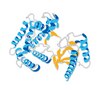
Keratin, type II cytoskeletal 8 (UniProt: P05787; also known as Cytokeratin-8, CK-8, Keratin-8, K8, Type-II keratin Kb8) is encoded by the KRT8 (also known as CYK8) gene (Gene ID: 3856) in human. Cytokeratins belong to a diverse group of intermediate filaments that participate in differentiation and maintain the structural integrity of epithelial cells. Cytokeratins share a common structural organization: a central alpha-helical rod domain flanked by head and tail domains. An important unique feature of cytokeratins is their epithelial cell-type-specific expression. CK8 is expressed in abundance in the epithelia of colon, bladder, ileum, and stomach. It is a type II, neutral to basic protein that together with cytokeratin-19 (KRT19) helps to link the contractile apparatus to dystrophin at the costameres of striated muscle. CK-8 can undergo phosphorylation on three major serine residues: Serine 23, 431, and 73. Serine 23 is shown to be highly conserved in all type II keratins. Phosphorylation at Serine 73 is reported to increase during cellular stress. However, under normal conditions serine 73 remains largely dephosphorylated. It can also undergo O-glycosylation in a cell cycle-dependent manner and glycosylation increases its solubility and reduces stability by inducing proteasomal degradation. CK8 expression has been correlated with malignancy in leukoplakia and carcinomas of the head and neck and its expression has been observed in all non-small-cell lung cancers.
References
Product Information
Format
Purified
Presentation
Purified mouse monoclonal antibody IgM in buffer containing 0.1 M Tris-Glycine (pH 7.4), 150 mM NaCl with 0.05% sodium azide.
Applications
Application
Anti-Cytokeratin 8, clone 35betaH11, Cat. No. MABT1529, is a mouse monoclonal antibody that detects Cytokeratin-8 and has been tested for use in Immunohistochemistry (Paraffin) and Western Blotting.
Key Applications
Immunohistochemistry (Paraffin)
Western Blotting
Application Notes
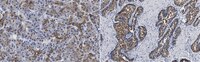
Immunohistochemistry (Paraffin) Analysis: A 1:250 dilution from a representative lot detected Cytokeratin-8 in human prostate tissue sections.
Western Blotting Analysis: A representative lot detected Cytokeratin-8 in Western Blotting applications (Gown, A.M., et. al. (1984). Am J Pathol. 114(2):309-21).
Immunohistochemistry (Paraffin) Analysis: A representative lot detected Cytokeratin-8 in Immunohistochemistry applications (Gown, A.M., et. al. (1984). Am J Pathol. 114(2):309-21).
Biological Information
Immunogen
A cytoskeletal preparation of the human hepatocellular carcinoma cell line Hep3B.
Clone
35betaH11
Concentration
Please refer to lot specific datasheet.
Host
Mouse
Specificity
Clone 35betaH11 is a mouse monoclonal antibody that specifically detetcts human Cytokeratin 8.
Evaluated by Immunohistochemistry (Paraffin) in human pancreas tissue sections.
Immunohistochemistry (Paraffin) Analysis: A 1:250 dilution of this antibody detected Cytokeratin-8 in human pancreas tissue sections.
Usage Statement
Unless otherwise stated in our catalog or other company documentation accompanying the product(s), our products are intended for research use only and are not to be used for any other purpose, which includes but is not limited to, unauthorized commercial uses, in vitro diagnostic uses, ex vivo or in vivo therapeutic uses or any type of consumption or application to humans or animals.
Monoclonal antibodies to human intermediate filament proteins. II. Distribution of filament proteins in normal human tissues. Gown, AM; Vogel, AM Am J Pathol
114
309-21
1983
Monoclonal antibodies generated against different human intermediate filament (IF) proteins were assayed on fixed, embedded tissue by the biotin-avidin-immunoperoxidase method for evaluation of the tissue specificity of these antibodies. An antibody (43 beta E8) made to fibroblast IF protein stains mesenchymal tissue such as endothelium, histiocytes, stromal fibroblasts, and Schwann cells but does not stain epithelium, skeletal muscle, lymphocytes, or neurons. Three different anti-cytokeratin antibodies decorate epithelium in three unique patterns. One (35 beta H11) stains all nonsquamous epithelium but fails to recognize squamous epithelium. Antibody 34 beta E12 stains the full thickness of squamous epithelium and ductular epithelium but does not react with hepatocytes, pancreatic acinar cells, proximal renal tubules, or endometrial glands. Antibody 34 beta B4 stains only the suprabasal portion of squamous epithelium. None of these three anti-cytokeratin antibodies reacts with nerve or mesenchymal tissue. Two anti-neurofilament antibodies recognize only neurons, failing to react with epithelial or mesenchymal tissue. We conclude that these anti-intermediate filament antibodies can be used as tissue-specific markers. Neoplasms retain the same intermediate filament patterns as the normal parental tissue; therefore, these antibodies can be used as diagnostic aids in surgical pathology.