


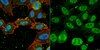
05-1139 Sigma-AldrichAnti-Focal Adhesion Kinase Antibody, clone BLAb2H7
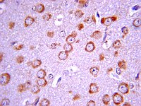


This Anti-Focal Adhesion Kinase Antibody, clone BLAb2H7 is validated for use in WB, IP for the detection of Focal Adhesion Kinase.
More>> This Anti-Focal Adhesion Kinase Antibody, clone BLAb2H7 is validated for use in WB, IP for the detection of Focal Adhesion Kinase. Less<<Anti-Focal Adhesion Kinase Antibody, clone BLAb2H7 : FDS (Fiches de données de sécurité), certificats d’analyse (CoA) et de qualité (CoQ), dossiers, brochures et autres documents disponibles.
Produits recommandés

Aperçu
| Replacement Information |
|---|
Tableau de caractéristiques principal
| Species Reactivity | Key Applications | Host | Format | Antibody Type |
|---|---|---|---|---|
| H, M, R, Ch | WB, IP | M | Purified | Monoclonal Antibody |
| References |
|---|
| Product Information | |
|---|---|
| Format | Purified |
| Control |
|
| Presentation | Purified mouse monoclonal IgG1 in buffer containing 0.05% sodium azide. |
| Quality Segment | MQ100 |
| Physicochemical Information |
|---|
| Dimensions |
|---|
| Materials Information |
|---|
| Toxicological Information |
|---|
| Safety Information according to GHS |
|---|
| Safety Information |
|---|
| Packaging Information | |
|---|---|
| Material Size | 100 µg |
| Transport Information |
|---|
| Supplemental Information |
|---|
| Specifications |
|---|
| Global Trade ITEM Number | |
|---|---|
| Référence | GTIN |
| 05-1139 | 04053252670091 |
Documentation
Required Licenses
| Title |
|---|
| PRODUCTO REGULADO POR LA SECRETARÍA DE SALUD |
Anti-Focal Adhesion Kinase Antibody, clone BLAb2H7 FDS
| Titre |
|---|