コンテンツへスキップ
Merck

MIDI67

PKH67緑色蛍光細胞リンカーMidiキット、一般細胞膜標識用

Distributed for Phanos Technologies

別名:

Green PKH membrane midi labeling kit

ログインで組織・契約価格をご覧ください。

サイズを選択してください


この商品について

NACRES:
NA.32
UNSPSC Code:
12352207
テクニカルサービス
お困りのことがあれば、経験豊富なテクニカルサービスチームがお客様をサポートします。
お手伝いします
テクニカルサービス
お困りのことがあれば、経験豊富なテクニカルサービスチームがお客様をサポートします。
お手伝いします

製品名

PKH67緑色蛍光細胞リンカーMidiキット、一般細胞膜標識用, Distributed for Phanos Technologies

packaging

pkg of 1 kit

manufacturer/tradename

Distributed for Phanos Technologies

storage condition

protect from light

technique(s)

flow cytometry: suitable

fluorescence

λex 490 nm; λem 502 nm (PKH67 dye)

application(s)

cell analysis
detection

detection method

fluorometric

shipped in

ambient

storage temp.

room temp

Quality Level

関連するカテゴリー

Application

PKH67緑色蛍光細胞リンカーMidiキットは以下の用途に用いられています:
  • エキソソームの標識  
  • 細胞外小胞(EV)
  • 正常および鎌状赤血球(RBC)
  • Raji細胞

General description

本キットは、幅広い細胞膜の標識に使用できます。PKH67は、in vitroおよびin vivoトラッキングで既述した他の2種の緑色色素PKH1およびPKH2より長い脂肪族炭素末端を有しています。尾部が長いことにより、社内試験ではPKH2と比較してPKH67では一貫して細胞-細胞伝達が遅いことが示されています。
PKH1およびPKH2を用いたin vivo試験で、蛍光の緩徐な消失が認められています。この挙動は緑色細胞リンカー色素の特性であり、赤色細胞リンカー色素の特性ではないと考えられるため、PKH67は同様の特性を示す可能性があります。 非分裂細胞でのin vitro細胞膜維持とin vivo蛍光半減期との相関から、PKH67のin vivo蛍光半減期は10~12日と予測されます。 同等の半減期を有する他の緑色細胞リンカー色素は、1~2か月の期間にわたってin vivoのリンパ球およびマクロファージのトラフィキングを調査するのに使用されており、PKH67は中程度の長さのin vivoトラッキング研究にも有用であることを示唆しています。

Other Notes

参考文献を含めPKH及びCellVue® Fluorescent Cell Linker Dyeのさらに詳しい情報につきましては、こちらをご覧ください。

Legal Information

CellVue is a registered trademark of Phanos Technologies

キットの構成要素のみ

製品番号
詳細

  • PKH67 Dye 2 x 0.1

  • Diluent C 6 x 10

pictograms

FlameExclamation mark

signalword

Danger

hcodes

Hazard Classifications

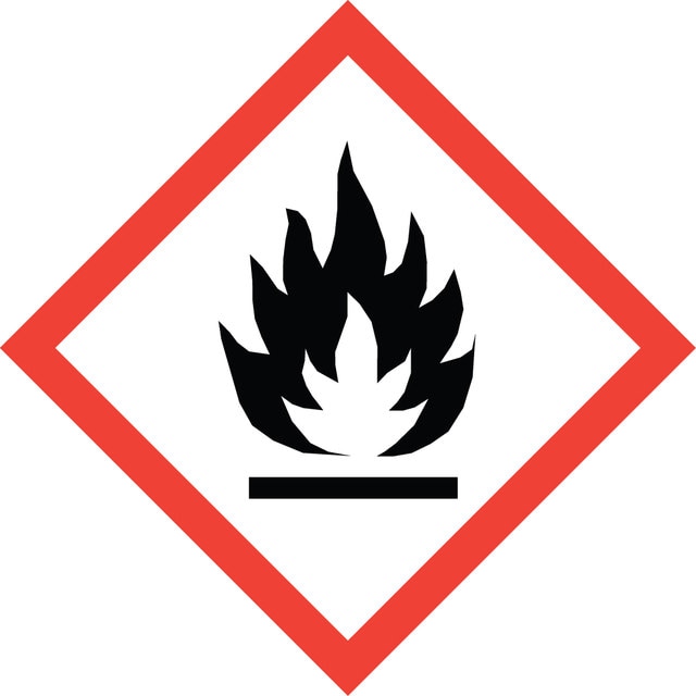
Eye Irrit. 2 - Flam. Liq. 2

保管分類

3 - Flammable liquids

flash_point_f

57.2 °F - closed cup

flash_point_c

14 °C - closed cup


適用法令

試験研究用途を考慮した関連法令を主に挙げております。化学物質以外については、一部の情報のみ提供しています。 製品を安全かつ合法的に使用することは、使用者の義務です。最新情報により修正される場合があります。WEBの反映には時間を要することがあるため、適宜SDSをご参照ください。

キットコンポーネントの情報を参照してください

pdsc

キットコンポーネントの情報を参照してください

prtr

キットコンポーネントの情報を参照してください

fsl

キットコンポーネントの情報を参照してください

ishl_indicated

キットコンポーネントの情報を参照してください

ishl_notified

キットコンポーネントの情報を参照してください

cart

キットコンポーネントの情報を参照してください

jan


最新バージョンのいずれかを選択してください:

試験成績書(COA)

Lot/Batch Number

適切なバージョンが見つかりませんか。

特定のバージョンが必要な場合は、ロット番号またはバッチ番号で特定の証明書を検索できます。

以前この製品を購入いただいたことがある場合

文書ライブラリで、最近購入した製品の文書を検索できます。

文書ライブラリにアクセスする

Dongxin Tang et al.
Clinical and translational medicine, 10(4), e139-e139 (2020-09-09)
Cancer stem cells (CSCs) are important factors contributing to tumorigenesis. We examined whether CSCs isolated from colorectal cancer (CRC) cells possess metastatic properties that can be transferred to non-CSCs via the delivery of miR-200c enclosed in extracellular vesicles (EVs). The
Notochordal-cell derived extracellular vesicles exert regenerative effects on canine and human nucleus pulposus cells
Bach F, et al.
Testing, 8(51), 88845-88845 (2017)
Modulation of Sickle Red Blood Cell Adhesion and its Associated Changes in Biomarkers by Sulfated Nonanticoagulant Heparin Derivative
Alshaiban A, et al.
Clinical and Applied Thrombosis/Hemostasis : Official Journal of the International Academy of Clinical and Applied Thrombosis/Hemostasis, 22, 230-238 (2016)
ADCC Assay Protocol
Fang L and He F
Journal of Virology (2013)
Ioulia Vardaki et al.
Oncotarget, 7(46), 74966-74978 (2016-09-03)
Breast cancer (BrCa) is the most frequent cancer type in women and a leading cause of cancer related deaths in the world. Despite the decrease in mortality due to better diagnostics and palliative care, there is a lack of prognostic

資料

Optimal staining is a key component for studying tumorigenesis and progression. Learn useful tips and techniques for dye applications, including examples from recent studies.

PKH and CellVue® Fluorescent Cell Linker Kits provide fluorescent labeling of live cells over an extended period of time, with no apparent toxic effects.

ライフサイエンス、有機合成、材料科学、クロマトグラフィー、分析など、あらゆる分野の研究に経験のあるメンバーがおります。.

製品に関するお問い合わせはこちら(テクニカルサービス)